. 2012 Aug;23(15):2891-904.
doi: 10.1091/mbc.E11-04-0383. Epub 2012 Jun 6.

Myosin 1E coordinates actin assembly and cargo trafficking during clathrin-mediated endocytosis

Affiliations

Myosin 1E coordinates actin assembly and cargo trafficking during clathrin-mediated endocytosis

Jackie Cheng et al. Mol Biol Cell. 2012 Aug.

Abstract

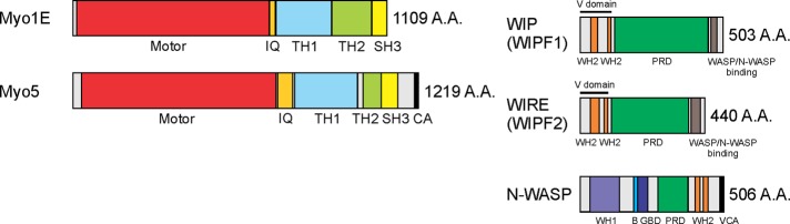
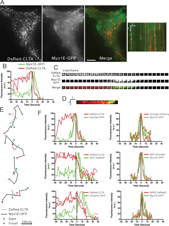
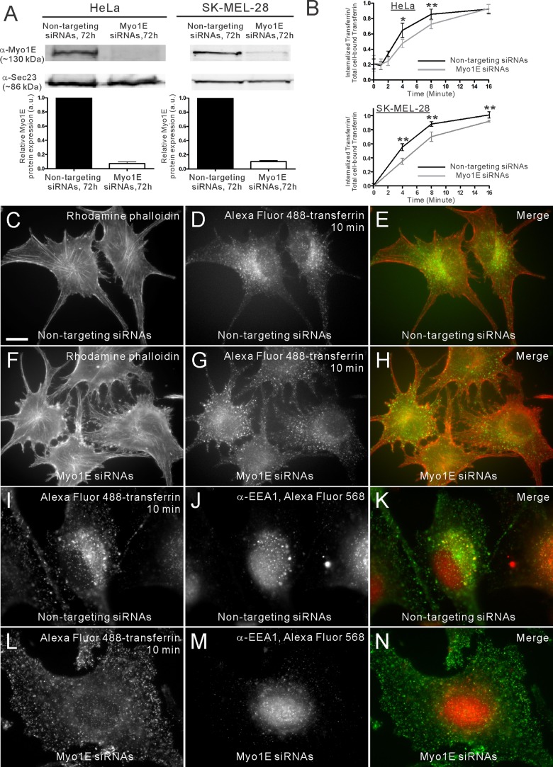
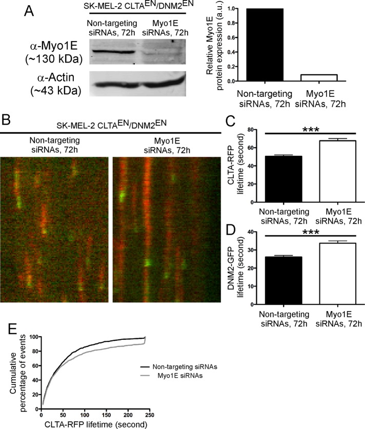
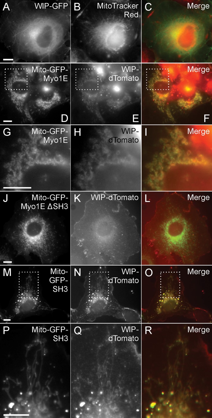
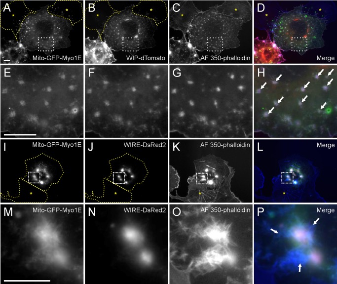
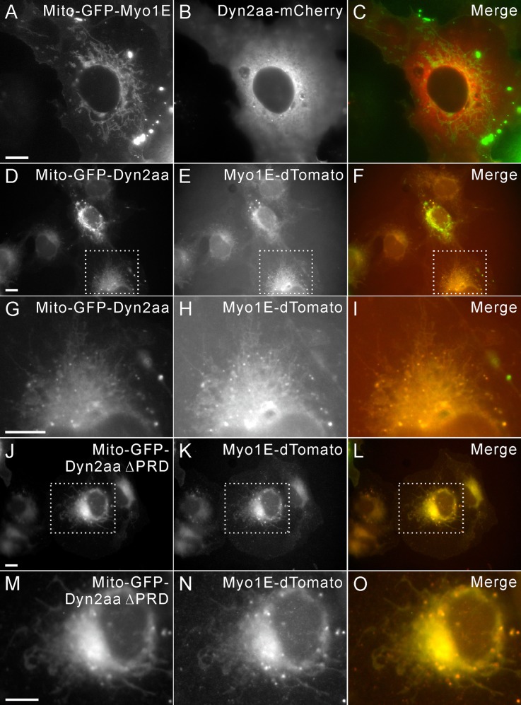
Myosin 1E (Myo1E) is recruited to sites of clathrin-mediated endocytosis coincident with a burst of actin assembly. The recruitment dynamics and lifetime of Myo1E are similar to those of tagged actin polymerization regulatory proteins. Like inhibition of actin assembly, depletion of Myo1E causes reduced transferrin endocytosis and a significant delay in transferrin trafficking to perinuclear compartments, demonstrating an integral role for Myo1E in these actin-mediated steps. Mistargeting of GFP-Myo1E or its src-homology 3 domain to mitochondria results in appearance of WIP, WIRE, N-WASP, and actin filaments at the mitochondria, providing evidence for Myo1E's role in actin assembly regulation. These results suggest for mammalian cells, similar to budding yeast, interdependence in the recruitment of type I myosins, WIP/WIRE, and N-WASP to endocytic sites for Arp2/3 complex activation to assemble F-actin as endocytic vesicles are being formed.


Figures

FIGURE 1:
FIGURE 1:
Schematic representation of the domain structure of mammalian type I myosin, Myo1E. Myo1E is composed of an N-terminal motor domain, a neck domain with IQ calmodulin-binding motif, a tail domain with tail-homology (TH) 1 and 2 domains, and a Src-homology (SH3) domain. Budding yeast Myo3/5 proteins have similar domain structures to the mammalian orthologue, Myo1E, except for the addition of a C-terminal (C) and acidic (A) domain. The A and C domains are necessary to activate the yeast Arp2/3 complex for actin polymerization. Mammalian WASP-interacting protein family 1/2 (WIPF1/2), commonly known as WIP and WIRE, related to yeast verprolin (Vrp1), is composed of two WASP-homology 2 (WH2) domains in the N-terminal verprolin-homology domain (V domain), a proline-rich domain (PRD), and a C-terminal WASP/N-WASP–binding domain. Mammalian N-WASP is composed of an N-terminal WIP-binding WASP-homology 1 (WH1) domain, a basic region (B), a GTPase-binding domain (GBD), a PRD, and a C-terminal VCA domain.
FIGURE 2:
FIGURE 2:
Recruitment of Myo1E-GFP when clathrin is being internalized. (A) A low-magnification TIRFM view of Swiss 3T3 cells stably expressing DsRed-clathrin light chain a (CLTA) and transiently expressing Myo1E-GFP. Right, a kymograph of the peripheral region of the same cell, showing recruitment of Myo1E to CCPs at the time of internalization. Bar, 10 μm. (B) Representative temporal recruitment profile of Myo1E-GFP and DsRed-CLTA in Swiss 3T3 cells. Myo1E-GFP is recruited concomitant with clathrin internalization. (C) Montage and (D) kymograph show the recruitment of Myo1E-GFP at the end of the lifetime of DsRed-CLTA. (E) Particle tracking of Myo1E-GFP shows a characteristic vectoral movement at the CCP with reference to CLTA. Bar, 100 nm. (F) Representative temporal recruitment profiles for GFP-tagged dynamin, N-WASP, and actin-marker utrophin (calponin-homology domain) in Swiss 3T3 cells stably expressing DsRed-CLTA (left). Images were captured at 2-s intervals. Representative temporal recruitment profiles of tagged dynamin, WIP, and WIRE in Swiss 3T3 cells expressing Myo1E-GFP (right). Images were captured at 1-s intervals.
FIGURE 3:
FIGURE 3:
Low levels of lat-A inhibit endocytic trafficking. (A–O) SK-MEL-28 cells were treated with either DMSO (A–C) or low levels of lat-A for 10 min and subjected to a fluorescent transferrin-uptake assay in the absence (D–F, J–L) or presence (G–I, M–O) of lat-A. (A–C) In the DMSO-treated controls, cells exhibit normal F-actin structure (A) and the Alexa Fluor 488–transferrin was internalized and trafficked to the perinuclear region (B). (C) The merge of A and B. (D–F) In cells treated with 0.05 μM lat-A, despite the mild disruption of F-actin (D), the transferrin uptake (E) is similar to the control (B). (F) The merge of D and E. (G–I) However, when these cells were incubated in the presence of lat-A during fluorescent transferrin-uptake assay, the endocytic trafficking of transferrin is inhibited (H). (I) The merge of G and H. (J–O) In cells treated with 0.1 μM lat-A, either in the absence (J–L) or presence (M–O) of lat-A during the transferrin-uptake assay, significant endocytic and trafficking defects were observed. (D, G, J, M) Cells treated with low levels of lat-A show continued presence of stress fibers. Bar, 10 μm.
FIGURE 4:
FIGURE 4:
Depletion of Myo1E reduces endocytosis and inhibits downstream endocytic trafficking. (A) Western blots and quantification of Myo1E depletion using siRNAs. Depletion of Myo1E in HeLa and SK-MEL-28 cells after 72 h showed a 96 and 90% reduction in Myo1E protein levels, respectively. Sec23 serves as a loading control. (B) ELISA-based transferrin-uptake assay showed a 27 and 36% reduction in internalization of biotinylated transferrin at 4-min in Myo1E-depleted HeLa and SK-MEL-28 cells compared with control, respectively. *p < 0.05 and **p < 0.01. (C, F) F-actin staining and (D, G) fluorescent transferrin uptake in control and Myo1E-depleted SK-MEL-28 cells, respectively. Ten minutes after internalization, Alexa Fluor 488-transferrin–containing puncta were concentrated at the perinuclear region in the control cells (D). (D, G) By quantifying the total fluorescence of the cell, there is a 51 ± 18% reduction of the internalized transferrin in Myo1E-depleted cells compared with the control cells (cells = 6 and 7 for Myo1E-depleted and control cells, respectively). In addition, the internalized puncta were localized at the cell periphery rather than at the perinuclear region. (I–K) Immunofluorescence shows colocalization of the perinuclear-concentrated Alexa Fluor 488–transferrin puncta and an early endosome marker EEA1 in the control cells. However, in the Myo1E-depleted cells (L–N), the puncta were dispersely localized and did not colocalize with EEA1, which retains its perinuclear localization. Bar, 10 μm.
FIGURE 5:
FIGURE 5:
Depletion of Myo1E in genome-edited CLTAEN/DNM2EN SK-MEL-2 cells increased the average lifetime of CLTA-RFP and DNM2-GFP. (A) Western blots and quantification of Myo1E depletion using siRNAs after 72 h showed a 91% reduction of Myo1E protein expression. (B) Representative examples of kymograph showing the lifetime of CLTA-RFP (red) and DNM2-GFP (green) in CLTAEN/DNM2EN cells treated with control or Myo1E siRNAs. (C) The average lifetime of CLTA-RFP in Myo1E-depleted cells (61.9 ± 2.4 s; tracks, 982; cells, 8) is significantly longer than that in the control cells (48.6 ± 2.1 s; tracks, 1293; cells, 8). (D) The average lifetime of DNM2-GFP in Myo1E-depleted cells (33.7 ± 1.3 s; tracks, 998; cells, 8) is also significantly longer than in the control cells (26.1 ± 0.85 s; tracks, 934; cells, 8). Data are presented as means ± SEM. ***p < 0.0001. (E) Cumulative percentage of frequencies of CLTA-RFP lifetime in the analysis.
FIGURE 6:
FIGURE 6:
Myo1E SH3 domain is necessary and sufficient to recruit WIP. (A–D) Transient expression of WIP-GFP (A) in Cos-7 control cells labeled with the mitochondria marker MitoTracker Red (B). WIP-GFP normally does not colocalize with MitoTracker Red. (C) The merge of A and B. (E, F) In cells cotransfected with mitochondria-targeted Mito-GFP-Myo1E and WIP-dTomato, WIP-dTomato is recruited to mitochondria. (G–I) High magnification of E and F showing the colocalization of GFP-Myo1E–decorated mitochondria and WIP-dTomato. (I) The merge of G and H. (J–R) SH3 domain of Myo1E is necessary and sufficient to recruit WIP-dTomato. (J–L) In cells cotransfected with Mito-GFP-Myo1E ΔSH3 and WIP-dTomato, WIP-dTomato is not recruited to the GFP-Myo1E-decorated mitochondria. (M–R) In cells cotransfected with Mito-GFP-SH3 and WIP-dTomato, the recruitment of WIP-dTomato is restored. (O) The merge of M and N. (P–R) High magnification of M–O shows the colocalization of GFP-SH3–decorated mitochondria and WIP-dTomato. (R) The merge of P and Q. Bars, 10 μm.
FIGURE 7:
FIGURE 7:
Actin assembly at the GFP-Myo1E and WIP-dTomato or WIRE-DsRed2-decorated mitochondria. Cotransfection of Mito-GFP-Myo1E and WIP-dTomato (A–H) or WIRE-DsRed2 (I–P) results in actin assembly at the mitochondria. Yellow asterisk marks an untransfected cell that has no aberrant actin structures in the cytoplasm. Arrows indicate actin meshwork assembled on the GFP-Myo1E and WIP-dTomato or WIRE-DsRed-decorated mitochondria. Bar, 10 μm.
FIGURE 8:
FIGURE 8:
Dynamin recruits Myo1E independent of its proline-rich domain. (A, B) Cotransfection of Mito-GFP-Myo1E and dynamin-mCherry shows that the latter is not recruited to the mitochondria. (C) The merge of A and B. (D–F) Cotransfection of Mito-GFP-dynamin and Myo1E-dTomato shows that Myo1E-dTomato is efficiently recruited to the GFP-dynamin–decorated mitochondria. (F) The merge of D and E. (G–I) High magnification of D–F showing the colocalization of GFP-dynamin–decorated mitochondria and Myo1E-dTomato. (I) The merge of G and H. (J–L) Cotransfection of Mito-GFP–dynamin ΔPRD and Myo1E-dTomato shows efficient recruitment of Myo1E-dTomato to the mitochondria. (L) The merge of J and K. (M–O) High magnification of J–L showing the colocalization of GFP-dynamin ΔPRD–decorated mitochondria and Myo1E-dTomato. (O) The merge of M and N. Bars, 10 μm.

Similar articles

Cited by

References

    1. Anderson BL, Boldogh I, Evangelista M, Boone C, Greene LA, Pon LA. The Src homology domain 3 (SH3) of a yeast type I myosin, Myo5p, binds to verprolin and is required for targeting to sites of actin polarization. J Cell Biol. 1998;141:1357–1370. - PMC - PubMed
    1. Ayscough KR, Stryker J, Pokala N, Sanders M, Crews P, Drubin DG. High rates of actin filament turnover in budding yeast and roles for actin in establishment and maintenance of cell polarity revealed using the actin inhibitor latrunculin-A. J Cell Biol. 1997;137:399–416. - PMC - PubMed
    1. Benesch S, Polo S, Lai FP, Anderson KI, Stradal TE, Wehland J, Rottner K. N-WASP deficiency impairs EGF internalization and actin assembly at clathrin-coated pits. J Cell Sci. 2005;118:3103–3115. - PubMed
    1. Boulant S, Kural C, Zeeh JC, Ubelmann F, Kirchhausen T. Actin dynamics counteract membrane tension during clathrin-mediated endocytosis. Nat Cell Biol. 2011;13:1124–1131. - PMC - PubMed
    1. Buss F, Arden SD, Lindsay M, Luzio JP, Kendrick-Jones J. Myosin VI isoform localized to clathrin-coated vesicles with a role in clathrin-mediated endocytosis. EMBO J. 2001;20:3676–3684. - PMC - PubMed

Publication types

LinkOut - more resources