. 2012 Sep;40(16):7896-906.
doi: 10.1093/nar/gks520. Epub 2012 Jun 6.

Re-splicing of mature mRNA in cancer cells promotes activation of distant weak alternative splice sites

Affiliations

Re-splicing of mature mRNA in cancer cells promotes activation of distant weak alternative splice sites

Toshiki Kameyama et al. Nucleic Acids Res. 2012 Sep.

Abstract

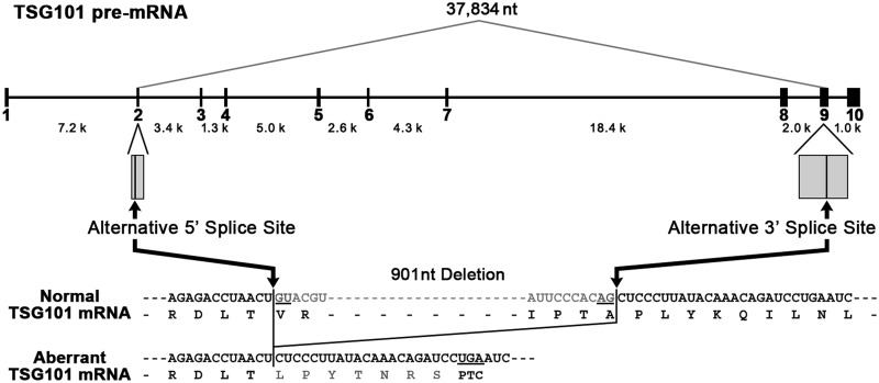
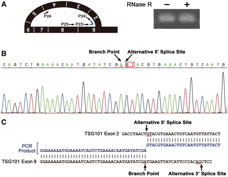
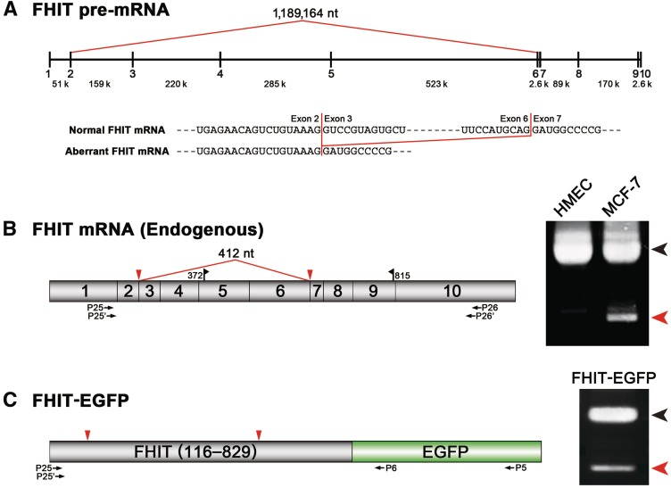
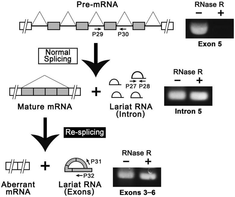
Transcripts of the human tumor susceptibility gene 101 (TSG101) are aberrantly spliced in many cancers. A major aberrant splicing event on the TSG101 pre-mRNA involves joining of distant alternative 5' and 3' splice sites within exon 2 and exon 9, respectively, resulting in the extensive elimination of the mRNA. The estimated strengths of the alternative splice sites are much lower than those of authentic splice sites. We observed that the equivalent aberrant mRNA could be generated from an intron-less TSG101 gene expressed ectopically in breast cancer cells. Remarkably, we identified a pathway-specific endogenous lariat RNA consisting solely of exonic sequences, predicted to be generated by a re-splicing between exon 2 and exon 9 on the spliced mRNA. Our results provide evidence for a two-step splicing pathway in which the initial constitutive splicing removes all 14 authentic splice sites, thereby bringing the weak alternative splice sites into close proximity. We also demonstrate that aberrant multiple-exon skipping of the fragile histidine triad (FHIT) pre-mRNA in cancer cells occurs via re-splicing of spliced FHIT mRNA. The re-splicing of mature mRNA can potentially generate mutation-independent diversity in cancer transcriptomes. Conversely, a mechanism may exist in normal cells to prevent potentially deleterious mRNA re-splicing events.


Figures

Figure 1.
Figure 1.
TSG101 pre-mRNA is aberrantly spliced in cancer cells by the activation of distant weak alternative splice sites. The structure of the TSG101 pre-mRNA and the major aberrant splicing often observed in various cancers are shown schematically. The sequences of the normal and aberrant TSG101 mRNAs are aligned with the encoded amino acids. The premature termination codon (PTC) generated in the aberrant mRNA is indicated.
Figure 2.
Figure 2.
Ectopically expressed intronless TSG101 gene (cDNA) is spliced and generates equivalent product as the endogenous aberrant mRNA in cancer cells. (A) The structure of the TSG101 mRNA. Red line and triangles indicate the postulated mRNA re-splicing via activated alternative 5′ and 3′ splice sites. Blue triangle indicates the PTC (1112–1114) generated by the postulated splicing. Black flags indicate the open reading frame (ORF; 127–1296). The positions of the PCR primers (P1–P4, P7–P10) are shown. (B) Using normal cells (HMEC) and cancer cells (MCF-7), endogenous TSG101 mRNAs were analyzed by RT-PCR (RT) and the genomic DNA was analyzed by PCR (Ge) with primer sets (P1–P4, P7–P10) annealed to the indicated exons. The black and red arrowheads denote the full-length and major aberrant mRNAs, respectively (Figure 1). (C) The structures of three reporter TSG101-EGFP plasmids (see (A) for the red and blue triangles). The postulated splicing-dependent EGFP expression of these plasmids is indicated with (−) and (+). The positions of the PCR primer sets (P1–P5, P3–P6) are shown. (D) Cancer cells (MCF-7) were transfected with the indicated TSG101-EGFP reporter plasmids and the EGFP expression was analyzed by fluorescence microscopy. (E) These ectopically expressed TSG101-EGFP transcripts were analyzed by RT-PCR. The black and red arrowheads indicate the unspliced and spliced products via the alternative splice sites, respectively. Two minor spliced products (indicated with * and **) were generated from different alternative 3′ splice sites, i.e. Δ190–776 (587-nt deletion) and Δ190–1236 (1047-nt deletion), respectively.
Figure 3.
Figure 3.
Detection of the lariat RNA consisting of only exons demonstrates the re-splicing of the endogenous TSG101 mRNA. (A) Schematic representation of the two postulated pathways leading to the generation of the aberrant mRNA, i.e. a conventional one-step direct aberrant splicing (right) and the proposed two-step process, including the re-splicing of the constitutively spliced mRNA (left). The pre-mRNA [1] and specific splicing products [2–5] from these two pathways were analyzed by RT-PCR with the indicated primers (arrows). (B) Detection of the specific splicing products [2–5] by RT-PCR using RNase R-digested (+) or RNase R-undigested (−) endogenous total RNA. The detected RT-PCR signals in the RNase R-digested sample (containing no linear RNAs) indicates an RNA species with either a 5′–2′ lariat or a 5′–3′ circular structure. However, the latter case was ruled out by the identification of a 5′–2′ branched structure (Figure 4A). Primers P19, P20, P21 and P22 anneal to introns 6, 7, 7 and 8, respectively. Primers P13/14 and P15/16 anneal to introns 7 and 8, respectively. Primers P11, P12, P17 and P18 anneal to exons 8, 10, 5 and 8, respectively. We used exactly the same PCR cycle numbers to amplify all these RNAs (Supplementary Materials and Methods).
Figure 4.
Figure 4.
The lariat RNA containing TSG101 exon 2 to exon 9 was identified by sequencing. (A) Detection of the lariat exons by lariat-specific RT-PCR amplification across the branch point with the indicated primer sets (P23–P24, P23′–P24′). The RT-PCR signal remained in the RNase R-digested (+) RNA sample, revealing a closed lariat structure. (B) Sequencing analysis of the lariat-specific RT-PCR product. Electropherogram of the sequence containing the branch point (arrow), followed by the alternative 5′ splice site (arrow) in the lariat exons. (C) A sequence alignment of the PCR fragment (blue) and the TSG101 gene (black) reveals a 2′–5′ branched connection between the end of the alternative 5′ splice site (GT) and the branch point (A), which is located upstream from the alternative 3′ splice site (AG).
Figure 5.
Figure 5.
Ectopically expressed intronless FHIT gene (cDNA) is spliced, generating a product equivalent to the endogenous aberrant mRNA in cancer cells. (A) Schematic representation of the FHIT pre-mRNA and the observed aberrant splicing in cancer cells (red line). The aligned sequences of the normal (full-length) and aberrant FHIT mRNAs reveal the skipping of exon 3 to exon 6. (B) Using normal cells (HMEC) and cancer cells (MCF-7), endogenous FHIT mRNAs (schematically shown) were analyzed by RT-PCR with the indicated primer sets (P25–P26, P25′–P26′). The full-length mRNA (black arrowhead) and aberrantly spliced mRNA (red arrowhead) are indicated. Black flags indicate the ORF (372–815) and the red line indicates the postulated mRNA re-splicing that skips the whole exon 3 to exon 6 region. (C) Cancer cells (MCF-7) were transfected with the FHIT-EGFP reporter plasmid (schematically shown) and the spliced products were analyzed by RT-PCR with the indicated primer sets (P25–P5, P25′–P6). The black and red arrowheads indicate the unspliced and spliced products, respectively. Sequence analysis mapped the spliced sites (red triangles) to the 5′ end of exon 3 and the 3′ end of exon 6 (data not shown).
Figure 6.
Figure 6.
Detection of a lariat RNA consisting only of exons demonstrates the re-splicing of the endogenous FHIT mRNA. RT-PCR analysis of the specific splicing products with the indicated primer sets (P27–P28, P29–P30, P31–P32) using RNase R-digested (+) or RNase R-undigested (−) endogenous total RNA. The RT-PCR signals in the RNase R-digested (+) sample (containing no linear RNAs) indicate either a lariat or circular structure, but a lariat structure was confirmed by the detection of the branched structure (Figure 7A). We used exactly the same number of PCR cycles to amplify all these RNAs (Supplementary Materials and Methods).
Figure 7.
Figure 7.
The lariat RNA containing FHIT exon 3 to exon 6 was identified by sequencing. (A) Detection of the lariat exons by a lariat-specific RT-PCR across the branch point with the indicated primer sets (P33–P34, P33′–P34′). The RT-PCR signal remained in the RNase R-digested (+) RNA sample, revealing a closed lariat structure. (B) The sequence of the RT-PCR product is shown in an electropherogram. The alternative 5′ splice site is indicated. (C) A sequence alignment of the PCR fragment (blue) with the FHIT gene (black) reveals a branched connection between the end of the alternative 5′ splice site (GT) and the branch point (A), which is located upstream from the alternative 3′ splice site (AG). A gap at the branch point (indicated with hyphens) results from skipping that occurred during reverse transcription, as described previously (48).
Figure 8.
Figure 8.
Different types of multi-step pre-mRNA splicing pathways. ‘Recursive splicing’ and ‘intrasplicing’ have been previously identified in Drosophila Ubx pre-mRNA and human EPB41 pre-mRNA, respectively (42,44,46). Novel multi-step splicing pathways were identified in human TSG101 and FHIT pre-mRNAs (right column), which are distinctive from the previously known multi-step splicing (left column). Each pathway of multi-step splicing is represented here with minimal numbers of exons/introns. 5′ and 3′ denote the 5′ splice site and the 3′ splice site, respectively. The black and gray colors indicate the active and inactive splice sites, respectively, in the each process.

Similar articles

Cited by

References

    1. Venables JP. Aberrant and alternative splicing in cancer. Cancer Res. 2004;64:7647–7654. - PubMed
    1. Kalnina Z, Zayakin P, Silina K, Line A. Alterations of pre-mRNA splicing in cancer. Genes Chromosomes Cancer. 2005;42:342–357. - PubMed
    1. Pajares MJ, Ezponda T, Catena R, Calvo A, Pio R, Montuenga LM. Alternative splicing: an emerging topic in molecular and clinical oncology. Lancet Oncol. 2007;8:349–357. - PubMed
    1. Li L, Cohen SN. Tsg101: a novel tumor susceptibility gene isolated by controlled homozygous functional knockout of allelic loci in mammalian cells. Cell. 1996;85:319–329. - PubMed
    1. Slagsvold T, Pattni K, Malerod L, Stenmark H. Endosomal and non-endosomal functions of ESCRT proteins. Trends Cell Biol. 2006;16:317–326. - PubMed

Publication types

MeSH terms