. 2012 Jul;139(14):2488-99.
doi: 10.1242/dev.078634. Epub 2012 Jun 6.

A Notch-dependent molecular circuitry initiates pancreatic endocrine and ductal cell differentiation

Affiliations

A Notch-dependent molecular circuitry initiates pancreatic endocrine and ductal cell differentiation

Hung Ping Shih et al. Development. 2012 Jul.

Abstract

In the pancreas, Notch signaling is thought to prevent cell differentiation, thereby maintaining progenitors in an undifferentiated state. Here, we show that Notch renders progenitors competent to differentiate into ductal and endocrine cells by inducing activators of cell differentiation. Notch signaling promotes the expression of Sox9, which cell-autonomously activates the pro-endocrine gene Ngn3. However, at high Notch activity endocrine differentiation is blocked, as Notch also induces expression of the Ngn3 repressor Hes1. At the transition from high to intermediate Notch activity, only Sox9, but not Hes1, is maintained, thus de-repressing Ngn3 and initiating endocrine differentiation. In the absence of Sox9 activity, endocrine and ductal cells fail to differentiate, resulting in polycystic ducts devoid of primary cilia. Although Sox9 is required for Ngn3 induction, endocrine differentiation necessitates subsequent Sox9 downregulation and evasion from Notch activity via cell-autonomous repression of Sox9 by Ngn3. If high Notch levels are maintained, endocrine progenitors retain Sox9 and undergo ductal fate conversion. Taken together, our findings establish a novel role for Notch in initiating both ductal and endocrine development and reveal that Notch does not function in an on-off mode, but that a gradient of Notch activity produces distinct cellular states during pancreas development.


Figures
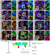
Fig. 1.
Fig. 1.
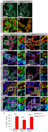
Sox9+ epithelial cord progenitors are Notch active. (A-L″) Immunofluorescence staining of mouse pancreas at E15.5 shows weak Notch1, but strong Notch2, and Rbpj expression in Hes1+ and Sox9+ cells of the progenitor epithelium (yellow arrowheads in A,B,D,E,H). Ngn3+ cells also express Notch2 and Rbpj (yellow arrowheads in F,I) but little Notch1 (white arrowheads in C). Hes1 is expressed in a subpopulation of Sox9+ cells that is Ngn3 (yellow arrowhead in J). Ngn3+ or Sox9+/Ngn3+ cells do not express Hes1 (white arrowheads in J,J′). The majority of Sox9+ cells express NICD1 and NICD2 (yellow arrowheads in K,L). β-Catenin (β-Cat), E-cadherin (Ecad) and DAPI visualize epithelial cells or cell nuclei. Insets show higher magnifications of boxed areas. (M) Graphic illustration of the Hes1, Sox9 and Ngn3 expression domains in the epithelial cords at E15.5. Ac, acinar; Endo, endocrine cluster; NICD, Notch intracellular domain. Scale bars: 40 μm.
Fig. 2.
Fig. 2.
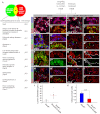
Notch signaling activates Hes1 and Sox9 at different thresholds. (A-H) Immunofluorescence staining of pancreatic explants from E12.5 Sox9-eGFP embryos cultured for 3 days in the presence of three different concentrations of γ-secretase inhibitor-IX (GSI-IX). At 1 μM GSI-IX, Hes1 expression is reduced, whereas Sox9 expression is maintained (B,F). At 5 μM and 10 μM GSI-IX, both Hes1 and Sox9 are severely diminished (C,D,G,H). (I) Quantitative RT-PCR analysis of Hes1 and Sox9 mRNA in explants cultured in different concentrations of GSI-IX (n=4). (J-M) Immunofluorescence staining of pancreata from Rosa26-NotchIC; Pdx1-Cre embryos at E15.5 demonstrates uniform Sox9 expression in NICD-expressing cells (marked by nGFP) (J). All NICD-expressing cells also express elevated level of Hes1 and a subset of targeted cells exhibit a Hes1high state (K). nGFP+ cells express the ductal marker Spp1 and DBA (M), but not Ngn3 (J), insulin (Ins) or amylase (AMY) (L). Insets show higher magnifications of boxed areas. (N) Graphical summary, correlating Notch activity with Hes1, Sox9 and Ngn3 expression. At high Notch activity, Hes1 and Sox9 are co-expressed, whereas intermediate Notch levels maintain Sox9, but not Hes1. β-Cat, β-catenin; DBA, Dolichos biflorus agglutinin. Values are shown as mean ± s.e.m. **P<0.01. Scale bars: 40 μm.
Fig. 3.
Fig. 3.
Sox9 cell-autonomously controls endocrine differentiation. (A-J) Immunofluorescence staining of E15.5 Sox9f/f (A,C,E,G), Sox9f/f; Rosa26-CreER (B,D,F,H), Ngn3+/eGFP; Sox9f/f (I) and Ngn3+/eGFP; Sox9f/f; Rosa26-CreER (J) embryos injected with high dosage of tamoxifen at E12.5. After Sox9 deletion, expression of Sox9 (B), Ngn3 protein (B) and Ngn3 mRNA (assessed by eGFP in J), endocrine hormones (D) and Nkx6.1 (H) are almost completely abolished. The acinar markers amylase (AMY) (F) and Ptf1a (H), or the ductal marker DBA (F) are not affected by Sox9 inactivation. E-cadherin (Ecad) visualizes epithelial cells. (K-R) Immunofluorescence staining of E15.5 Rosa26-eYFP; Sox9+/+; Pdx1-CreER (K,M,O,Q) and Rosa26-eYFP; Sox9f/f; Pdx1-CreER (L,N,P,R) embryos injected with low dosage of tamoxifen for mosaic Sox9 deletion. Recombined cells are traced by eYFP. (S) Quantification of marker+/eYFP+ cells relative to the total number of marker+ cells (n=3). Sox9-deficient cells show reduced propensity to differentiate into endocrine cells. Endo, all four endocrine hormones; DBA, Dolichos biflorus agglutinin. Values are shown as mean ± s.e.m. *P<0.05, **P<0.01. Scale bars: 40 μm.
Fig. 4.
Fig. 4.
Ngn3 cell-autonomously represses Sox9. (A-C‴) Immunofluorescence staining of Pdx1-Ngn3ER-IRES-nGFP embryos injected with tamoxifen at E8.75 (B-B‴) or E11.75 (C-C‴) and analyzed at E9.5 or E13.5, respectively. Tamoxifen-mediated nuclear translocation of the Ngn3-ER™ protein results in rapid and significant reduction of Sox9 (B-C‴). Dashed line outlines the pancreatic epithelium. (D-F) Transient transfection of HepG2 cells reveals robust Sox9 expression in pCMV-IRES-eGFP- (yellow arrowhead in D), but not in pCMV-Ngn3-IRES-eGFP-transfected cells (white arrowhead in E). (F) Quantification of eGFP+/Sox9+ cells relative to the total number of eGFP+ cells (n=3). Values are shown as mean ± s.e.m. **P<0.01. Scale bars: 40 μm.
Fig. 5.
Fig. 5.
Ngn3 deficiency results in reduced Ngn3 promoter activity and ectopic expression of ductal markers. (A-D) Whole-mount fluorescence image of Ngn3eGFP/eGFP and Ngn3+/eGFP embryos shows reduced eGFP signal in Ngn3-deficient embryos at E15.5. The pancreas is outlined by a dashed line (A,B). In Ngn3-deficient embryos, GFP+ cells retain E-cadherin (Ecad) expression (D), suggesting failure to delaminate from the epithelial cords. (E-N″) Immunofluorescence staining for progenitor and ductal cell markers on pancreatic sections from Ngn3+/eGFP (E,G,I,K,M) and Ngn3eGFP/eGFP (F,H,J,L,N) embryos at E15.5. eGFP+ cells in Ngn3+/eGFP pancreata are largely devoid of Sox9, Hes1, Tcf2, Prox1 and Spp1 (E,G,I,M), whereas eGFP+ cells in Ngn3eGFP/eGFP embryos retain expression of these markers (F,H,J,N). The pre-acinar marker Ptf1a is not detected in eGFP+ cells of either Ngn3+/GFP or Ngn3eGFP/eGFP embryos (K,L). Insets show higher magnifications of boxed areas. (O) Quantification of marker+/eGFP+ cells relative to the total number of eGFP+ cells (n=3). Du, duodenum. Values are shown as mean ± s.e.m. **P<0.01. Scale bars: 40 μm.
Fig. 6.
Fig. 6.
Sox9 is required for establishing a ductal epithelium. (A) Sox9-regulated genes were identified by comparing mRNA profiles of E15.5 pancreata from Sox9f/f; Rosa26-CreER and Sox9f/f littermates after inducing Sox9-deletion by tamoxifen injection at E12.5 (>1.5-fold change; n=4) and duct-enriched genes by comparing eGFP+ and eGFP- populations in Sox9-eGFP mice at p23 (n=3). The ten genes exhibiting a >2-fold decrease in Sox9-deficient embryos are listed. (B-I″) Immunofluorescence staining for ductal cell markers on pancreatic sections of Sox9f/f (B,D,F,H) and Sox9f/f; Rosa26-CreER (C,E,G,I) embryos injected with tamoxifen at E12.5 and analyzed at E15.5. Sox9 deficiency results in dilated lumens (asterisks in C,E,G,I). Expression of apical markers mucin 1 (Muc1), prominin 1 (CD133) (C) and ZO-1 (I) are not affected by Sox9 deletion. However, Sox9 inactivation results in loss of apically secreted Spp1 (E), polycystic kidney disease 2 (Pkd2; G) and the primary cilia marker acetylated-α-tubulin (AcTUB; I). E-cadherin (Ecad) visualizes epithelial cells. (J,K) Quantification of lumen diameter and ciliated luminal cells at E15.5 (n=3). Values are shown as mean ± s.e.m. **P<0.01. Scale bars: 40 μm.
Fig. 7.
Fig. 7.
High Notch activity in the absence of Sox9 does not restore endocrine or ductal cell differentiation. (A-R″) Immunofluorescence staining for ductal, endocrine and acinar markers on E15.5 pancreatic sections from Rosa26-eYFP; Ngn3-Cre (A,D,G,J,M,P), Rosa26-NotchIC; Ngn3-Cre (B,E,H,K,N,Q) and Rosa26-NotchIC; Sox9f/f; Ngn3-Cre (C,F,I,L,O,R) embryos. In Rosa26-eYFP; Ngn3-Cre embryos, the majority of targeted cells, traced by eYFP, express insulin (Ins), but not ductal or acinar markers. Activation of NICD in Ngn3-expressing cells forces cells to express ductal markers and inhibits endocrine or acinar differentiation. Deletion of Sox9 in NICD+ cells results in loss of ductal markers, Spp1, Pkd2, DBA, acetylated-α-tubulin (AcTUB) (L,O,R) and fails to restore endocrine differentiation (C,I). Insets show higher magnifications of boxed areas and E-cadherin (Ecad) visualizes epithelial cells. (S) Our data support a model whereby Notch signaling induces inhibitors and activators of endocrine differentiation at different thresholds. At high Notch activity Hes1-mediated Ngn3 repression prevents Sox9 from inducing Ngn3. At intermediate Notch levels, Hes1 is not maintained, allowing Sox9 to enable a self-promoting molecular circuitry that drives endocrine differentiation. Negative feedback of Ngn3 on Sox9 ensures exit from the bipotential ductal/endocrine state and terminal endocrine commitment. Cells remaining Sox9+/Hes1+ will acquire a ductal fate. Lu, lumen; DBA, Dolichos biflorus agglutinin. Scale bars: 40 μm.

Similar articles

Cited by

References

    1. Ahnfelt-Ronne J., Hald J., Bodker A., Yassin H., Serup P., Hecksher-Sorensen J. (2007). Preservation of proliferating pancreatic progenitor cells by Delta-Notch signaling in the embryonic chicken pancreas. BMC Dev. Biol. 7, 63 - PMC - PubMed
    1. Ahnfelt-Ronne J., Jorgensen M. C., Klinck R., Jensen J. N., Fuchtbauer E. M., Deering T., Macdonald R. J., Wright C. V., Madsen O. D., Serup P. (2011). Ptf1a-mediated control of Dll1 reveals an alternative to the lateral inhibition mechanism. Development 139, 33-45 - PMC - PubMed
    1. Akiyama H., Chaboissier M. C., Martin J. F., Schedl A., de Crombrugghe B. (2002). The transcription factor Sox9 has essential roles in successive steps of the chondrocyte differentiation pathway and is required for expression of Sox5 and Sox6. Genes Dev. 16, 2813-2828 - PMC - PubMed
    1. Apelqvist A., Li H., Sommer L., Beatus P., Anderson D. J., Honjo T., Hrabe de Angelis M., Lendahl U., Edlund H. (1999). Notch signalling controls pancreatic cell differentiation. Nature 400, 877-881 - PubMed
    1. Beucher A., Martin M., Spenle C., Poulet M., Collin C., Gradwohl G. (2011). Competence of failed endocrine progenitors to give rise to acinar but not ductal cells is restricted to early pancreas development. Dev. Biol. 361, 277-285 - PMC - PubMed

Publication types

MeSH terms