

Mast cell adenosine receptors function: a focus on the a3 adenosine receptor and inflammation
- PMID: 22675325
- PMCID: PMC3366457
- DOI: 10.3389/fimmu.2012.00134
Mast cell adenosine receptors function: a focus on the a3 adenosine receptor and inflammation
Abstract
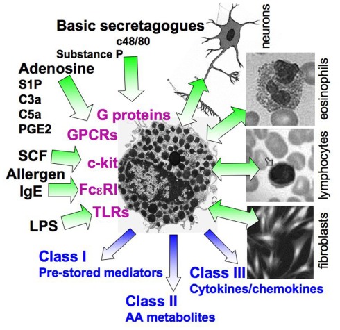
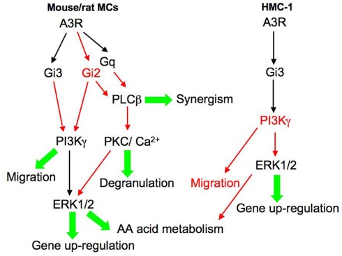
Adenosine is a metabolite, which has long been implicated in a variety of inflammatory processes. Inhaled adenosine provokes bronchoconstriction in asthmatics or chronic obstructive pulmonary disease patients, but not in non-asthmatics. This hyper responsiveness to adenosine appears to be mediated by mast cell activation. These observations have marked the receptor that mediates the bronchoconstrictor effect of adenosine on mast cells (MCs), as an attractive drug candidate. Four subtypes (A1, A2a, A2b, and A3) of adenosine receptors have been cloned and shown to display distinct tissue distributions and functions. Animal models have firmly established the ultimate role of the A3 adenosine receptor (A3R) in mediating hyper responsiveness to adenosine in MCs, although the influence of the A2b adenosine receptor was confirmed as well. In contrast, studies of the A3R in humans have been controversial. In this review, we summarize data on the role of different adenosine receptors in mast cell regulation of inflammation and pathology, with a focus on the common and distinct functions of the A3R in rodent and human MCs. The relevance of mouse studies to the human is discussed.
Keywords: A3 adenosine receptor; HMC-1; RBL-2H3; adenosine; mast cells.
Figures
Similar articles
-
Targeting adenosine receptors: novel therapeutic targets in asthma and chronic obstructive pulmonary disease.Am J Respir Med. 2002;1(2):99-105. doi: 10.1007/BF03256599. Am J Respir Med. 2002. PMID: 14720064 Review.
-
Mast cell-mediated stimulation of angiogenesis: cooperative interaction between A2B and A3 adenosine receptors.Circ Res. 2003 Mar 21;92(5):485-92. doi: 10.1161/01.RES.0000061572.10929.2D. Epub 2003 Feb 13. Circ Res. 2003. PMID: 12600879
-
Down-regulation of the A3 adenosine receptor in human mast cells upregulates mediators of angiogenesis and remodeling.Mol Immunol. 2015 May;65(1):25-33. doi: 10.1016/j.molimm.2014.12.015. Epub 2015 Jan 16. Mol Immunol. 2015. PMID: 25597247
-
A3 adenosine receptor activation triggers phosphorylation of protein kinase B and protects rat basophilic leukemia 2H3 mast cells from apoptosis.Mol Pharmacol. 2001 Jan;59(1):76-82. doi: 10.1124/mol.59.1.76. Mol Pharmacol. 2001. PMID: 11125027
-
Adenosine A2B receptors: a novel therapeutic target in asthma?Trends Pharmacol Sci. 1998 Apr;19(4):148-53. doi: 10.1016/s0165-6147(98)01179-1. Trends Pharmacol Sci. 1998. PMID: 9612090 Review.
Cited by
-
Role of Mast Cell-Derived Adenosine in Cancer.Int J Mol Sci. 2019 May 27;20(10):2603. doi: 10.3390/ijms20102603. Int J Mol Sci. 2019. PMID: 31137883 Free PMC article. Review.
-
Adenosine Signaling in Autoimmune Disorders.Pharmaceuticals (Basel). 2020 Sep 22;13(9):260. doi: 10.3390/ph13090260. Pharmaceuticals (Basel). 2020. PMID: 32971792 Free PMC article. Review.
-
Hypothermia in mouse is caused by adenosine A1 and A3 receptor agonists and AMP via three distinct mechanisms.Neuropharmacology. 2017 Mar 1;114:101-113. doi: 10.1016/j.neuropharm.2016.11.026. Epub 2016 Nov 30. Neuropharmacology. 2017. PMID: 27914963 Free PMC article.
-
Rapid Mast Cell Generation from Gata2 Reporter Pluripotent Stem Cells.Stem Cell Reports. 2018 Oct 9;11(4):1009-1020. doi: 10.1016/j.stemcr.2018.08.007. Epub 2018 Sep 6. Stem Cell Reports. 2018. PMID: 30197119 Free PMC article.
-
Potential effector and immunoregulatory functions of mast cells in mucosal immunity.Mucosal Immunol. 2015 May;8(3):444-63. doi: 10.1038/mi.2014.131. Epub 2015 Feb 11. Mucosal Immunol. 2015. PMID: 25669149 Free PMC article. Review.
References
-
- Ali H., Choi O. H., Fraundorfer P. F., Yamada K., Gonzaga H. M., Beaven M. A. (1996). Sustained activation of phospholipase D via adenosine A3 receptors is associated with enhancement of antigen- and Ca2+-ionophore-induced secretion in a rat mast cell line. J. Pharmacol. Exp. Ther. 276, 837–845 - PubMed
LinkOut - more resources
Full Text Sources
Other Literature Sources