. 2012;7(6):e38115.
doi: 10.1371/journal.pone.0038115. Epub 2012 Jun 4.

Real-time self-regulation of emotion networks in patients with depression

Affiliations

Real-time self-regulation of emotion networks in patients with depression

David E J Linden et al. PLoS One. 2012.

Abstract

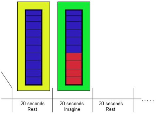
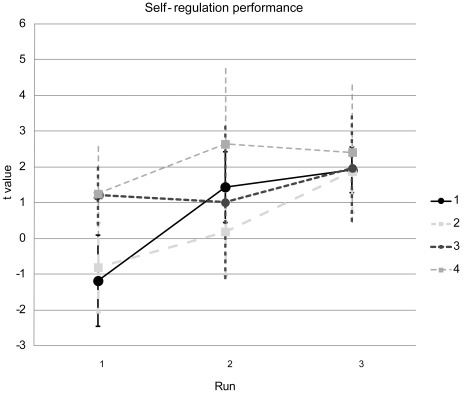
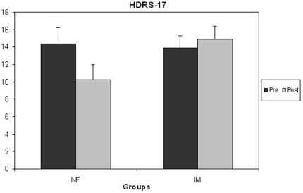
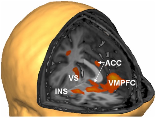
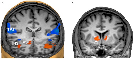
Many patients show no or incomplete responses to current pharmacological or psychological therapies for depression. Here we explored the feasibility of a new brain self-regulation technique that integrates psychological and neurobiological approaches through neurofeedback with functional magnetic resonance imaging (fMRI). In a proof-of-concept study, eight patients with depression learned to upregulate brain areas involved in the generation of positive emotions (such as the ventrolateral prefrontal cortex (VLPFC) and insula) during four neurofeedback sessions. Their clinical symptoms, as assessed with the 17-item Hamilton Rating Scale for Depression (HDRS), improved significantly. A control group that underwent a training procedure with the same cognitive strategies but without neurofeedback did not improve clinically. Randomised blinded clinical trials are now needed to exclude possible placebo effects and to determine whether fMRI-based neurofeedback might become a useful adjunct to current therapies for depression.


Conflict of interest statement

Competing Interests: The authors have declared that no competing interests exist.

Figures

Figure 1
Figure 1. Neurofeedback protocol.
During the neurofeedback runs (3 in each of the 4 sessions), participants alternated between 20 s periods of rest and 20 s periods where they had to upregulate activity in the target area. The level of activation was fed back in real time (updated for each TR of 2 s) through the thermometer display.
Figure 2
Figure 2. Neurofeedback success.
Although self-regulation performance was varied during the first run (indicated by the low t values, scaled on the y-axis), participants achieved reliable upregulation during runs 2 and 3, with more stability in the later sessions. Data points represent group means and error bars represent the SD.
Figure 3
Figure 3. Neurofeedback produced clinical improvement that was not seen in the control group.
Patients in the neurofeedback (NF) treatment group, but not those in the imagery (IM) control group, improved significantly on the 17-item Hamilton Depression Rating scale, a standard clinical measure of depression severity and treatment effects. Lower values denote clinical improvement (error bars: standard errors).
Figure 4
Figure 4. The localiser procedure identified networks of positive mood.
Higher activation of right insula (INS), ventral striatum (VS), anterior cingulate cortex (ACC) and ventromedial prefrontal cortex (VMPFC) during presentation of positive compared to neutral images in the localiser runs (for full list of areas see Table 2). The localiser runs were effective in identifying brain areas responsive to positive images, which were used as target regions of interests (ROIs) for the subsequent neurofeedback procedure. The figure shows the contrast map thresholded at p<.05 (cluster level corrected) on a sample brain seen from the right and front (Talairach coordinates of virtual cuts: x = 0, y = 0, z = −2).
Figure 5
Figure 5. Network activation and deactivation during neurofeedback.
a) Activation of the insular cortex (INS) bilaterally and the right ventral striatum (VS) supported the neurofeedback task, whereas the temporoparietal junctions (TPJ) of both hemispheres were deactivated. The TPJ is recognised as part of the brain’s “default mode network” that is deactivated during effortful tasks. For a full documentation of the activated and deactivated networks see Table 3. View from the front and above. The right side of the brain is on the observer’s left (Talairach coordinates of virtual cuts: y = 25, z = −2). b) Successive training sessions produced further increases of activation during upregulation periods in the VS bilaterally (coronal view at y = 7, the right side of the brain is on the observer’s left).

References

    1. Kessler R, Berglund P, Demler O, Jin R, Merikangas K, et al. Lifetime prevalence and age-of-onset distributions of DSM-IV disorders in the National Comorbidity Survey Replication. Arch Gen Psychiatry. 2005;62:593–602. - PubMed
    1. Rush A, Trivedi M, Wisniewski S, Nierenberg A, Stewart J, et al. Acute and longer-term outcomes in depressed outpatients requiring one or several treatment steps: a STAR*D report. Am J Psychiatry. 2006;163:1905–17. - PubMed
    1. Mayberg H, Lozano A, Voon V, McNeely H, Seminowicz D, et al. Deep brain stimulation for treatment-resistant depression. Neuron. 2005;45:651–60. - PubMed
    1. George M, Aston-Jones G. Noninvasive techniques for probing neurocircuitry and treating illness: vagus nerve stimulation (VNS), transcranial magnetic stimulation (TMS) and transcranial direct current stimulation (tDCS). Neuropsychopharmacology. 2010;35:301–16. - PMC - PubMed
    1. Clark D, Beck A. Cognitive theory and therapy of anxiety and depression: convergence with neurobiological findings. Trends Cogn Sci. 2010;14(9):418–24. - PubMed

Publication types