. 2012;7(6):e38488.
doi: 10.1371/journal.pone.0038488. Epub 2012 Jun 4.

Networked T cell death following macrophage infection by Mycobacterium tuberculosis

Affiliations

Networked T cell death following macrophage infection by Mycobacterium tuberculosis

Stephen H-F Macdonald et al. PLoS One. 2012.

Abstract

Background: Depletion of T cells following infection by Mycobacterium tuberculosis (Mtb) impairs disease resolution, and interferes with clinical test performance that relies on cell-mediated immunity. A number of mechanisms contribute to this T cell suppression, such as activation-induced death and trafficking of T cells out of the peripheral circulation and into the diseased lungs. The extent to which Mtb infection of human macrophages affects T cell viability however, is not well characterised.

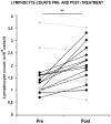
Methodology/principal findings: We found that lymphopenia (<1.5 × 10(9) cells/l) was prevalent among culture-positive tuberculosis patients, and lymphocyte counts significantly improved post-therapy. We previously reported that Mtb-infected human macrophages resulted in death of infected and uninfected bystander macrophages. In the current study, we sought to examine the influence of infected human alveolar macrophages on T cells. We infected primary human alveolar macrophages (the primary host cell for Mtb) or PMA-differentiated THP-1 cells with Mtb H37Ra, then prepared cell-free supernatants. The supernatants of Mtb-infected macrophages caused dose-dependent, caspase-dependent, T cell apoptosis. This toxic effect of infected macrophage secreted factors did not require TNF-α or Fas. The supernatant cytotoxic signal(s) were heat-labile and greater than 50 kDa in molecular size. Although ESAT-6 was toxic to T cells, other Mtb-secreted factors tested did not influence T cell viability; nor did macrophage-free Mtb bacilli or broth from Mtb cultures. Furthermore, supernatants from Mycobacterium bovis Bacille de Calmette et Guerin (BCG)- infected macrophages also elicited T cell death suggesting that ESAT-6 itself, although cytotoxic, was not the principal mediator of T cell death in our system.

Conclusions: Mtb-Infected macrophages secrete heat-labile factors that are toxic to T cells, and may contribute to the immunosuppression seen in tuberculosis as well as interfere with microbial eradication in the granuloma.


Conflict of interest statement

Competing Interests: The authors have declared that no competing interests exist.

Figures

Figure 1
Figure 1. Patient lymphocyte counts pre- and post antitubercular chemotherapy.
Lymphocyte counts were taken, before and after treatment, from twenty patients who were culture-positive for Mtb. Each joined pair of data points represents pre- and post-therapy lymphocyte counts (×109 cells/l) from a single patient. Data from patients whose lymphocyte counts recovered following treatment are shown in black; counts from patients whose lymphocyte counts fell or did not change are shown in grey. ** =  p<0.01, paired Student’s t test comparing pre-treatment vs post-treatment counts.
Figure 2
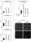
Figure 2. M.tuberculosis-infected macrophage supernatants are toxic to T cells.
(A) Jurkat T cells underwent significantly increased cell death in cell-free supernatants of low MOI and high MOI Mtb-infected human primary alveolar macrophages (AMs) (shaded bars; infection + and ++, respectively), compared to those of uninfected AMs (closed bars, infection –). Staurosporine 1 µM was used as a positive control to induce cell death (open bars). Similar effects were observed on PBTL and Jurkat T cells exposed to the cell-free supernatants of uninfected/infected THP-1 macrophages. (B) Jurkat cell death in infected THP-1 macrophage supernatant occurred in a dose-dependent manner; infected macrophage supernatants were diluted to 50% and 20% in uninfected supernatant, reducing the level of T cell killing. (C) Example cropped fluorescence intensity images for Hoechst and PI staining from Jurkats incubated in uninfected and infected AM supernatants. Images were processed using automated INCell image analysis to detect apoptotic condensed and fragmented nuclei (arrow 1 and 2, respectively) and PI-positive cells (arrow head). Data in each panel are from one representative experiment, showing mean percentage cell death ± SEM from incubations performed in triplicate. ** =  p<0.01, *** =  p<0.001, Student’s t test relative to uninfected control.
Figure 3
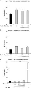
Figure 3. Macrophage-free M.tuberculosis bacilli are not sufficient to induce T cell death.
PBTLs (A) and Jurkat T cells (B) were incubated either in complete RPMI only (closed bars) or coincubated with Mtb bacilli at a range of T cell: Mtb ratios (shaded bars). Subsequently, Jurkat T cells were incubated in complete RPMI supplemented with medium taken from Mtb growing in the log phase at a range of ratios (shaded bars) (C). Staurosporine 1 µM was used as a positive control to induce cell death. Data in each panel are from one representative experiment, showing mean percentage cell death ± SEM from incubations performed in triplicate. *** =  p<0.001, Student’s t test relative to uninfected control.
Figure 4
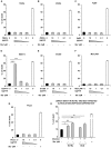
Figure 4. T cell death after exposure to M.tuberculosis secreted antigens.
Jurkat T cells were exposed for 24 h to a panel of secreted factors derived from Mtb. No increase in cell death occurred when T cells were incubated with 16 kDa, 45 kDa, Antigen 85 complex, GroES, ManLAM or Phos1 (A–C, E–G, respectively, shaded bars), however elevated death occurred in cells incubated with ESAT-6 at 10 µg/ml or 1 µg/ml (D). 1 µM Staurosporine was used as a positive control in each case (open bars); vehicle control was either PBS or DMSO in the case of ManLAM (closed bars). Supernatants of AMs infected with H37Ra or M.bovis BCG both significantly increased T cell death (H, shaded bars), compared with uninfected AM supernatants (H, closed bars). Data in each panel are from one representative experiment, showing mean percentage cell death ± SEM from incubations performed in triplicate. * =  p<0.05, ** =  p<0.01, *** =  p<0.001, Student’s t test relative to vehicle (A–G) or uninfected (H) control.
Figure 5
Figure 5. T cell death in M.tuberculosis-infected macrophage supernatants is caspase-dependent, and independent of TNF-α and Fas.
(A) Addition of the pan-caspase inhibitor Z-VAD-FMK mitigated cell death induced by low MOI and high MOI-infected macrophage supernatants (shaded bars, infection + and ++ respectively), as well as background cell death observed in the uninfected control supernatants (closed bars, infection -). Cell death in infected supernatants was not mitigated by coincubation with blocking antibodies to TNF-α (B) or Fas (C), and in each case was significantly higher than in uninfected control supernatants (closed bars). Cell death induced by positive controls (D) (1 µM Staurosporine, 5 ng/ml recombinant human TNF-α +0.2 µg/ml cycloheximide and 10 ng/ml recombinant human Fas ligand) was in each case significantly reduced by coincubation with the corresponding inhibitor (open bars). Data shown in each panel are from one representative experiment, showing mean percentage cell death ± SEM from triplicate incubations. ** =  p<0.01, *** =  p<0.001, Student’s t test relative to uninfected control (A–C), or positive control relative to inhibitor (D).
Figure 6
Figure 6. T cell death in M.tuberculosis-infected macrophage culture supernatants is mediated by heat-sensitive factor(s) greater than 50 kDa in molecular size.
(A) Heat inactivation (95°C, 20 min) of supernatants from low MOI and high MOI-infected macrophages (shaded bars, infection + and ++ respectively) inhibited induction of Jurkat T cell death. (B) Death-inducing factor(s) in infected macrophage supernatants were retained in the >50 kDa fractions following molecular size separation, and killed Jurkat T cells in a dose-dependent manner (25% and 10% dilution). Data shown in each panel are from one representative experiment, showing fold-change in cell death ± SEM in low MOI and high MOI-infected macrophage supernatants (shaded bars, infection + and ++, respectively) relative to uninfected control (closed bars), from incubations performed in triplicate. Each experiment was repeated a minimum of three times. ** =  p<0.01, *** =  p<0.001, Student’s t test relative to uninfected control.

References

    1. Davoudi S, Rasoolinegad M, Younesian M, Hajiabdolbaghi M, Soudbakhsh A, et al. CD4+ cell counts in patients with different clinical manifestations of tuberculosis. Brazilian Journal of Infectious Diseases. 2008;12:483–486. - PubMed
    1. Jones BE, Oo MM, Taikwel EK, Qian D, Kumar A, et al. CD4 Cell Counts in Human Immunodeficiency Virus-Negative Patients with Tuberculosis. Clinical Infectious Diseases. 1997;24:988–991. - PubMed
    1. Baena A, Porcelli SA. Evasion and subversion of antigen presentation by Mycobacterium tuberculosis. Tissue Antigens. 2009;74:189–204. - PMC - PubMed
    1. Schmolke M, García-Sastre A. Evasion of innate and adaptive immune responses by influenza A virus. Cellular Microbiology. 2010;12:873–880. - PMC - PubMed
    1. Gutierrez MG, Master SS, Singh SB, Taylor GA, Colombo MI, et al. Autophagy Is a Defense Mechanism Inhibiting BCG and Mycobacterium tuberculosis Survival in Infected Macrophages. Cell. 2004;119:753–766. - PubMed

Publication types

MeSH terms