. 2012 Jun;2(6):a006411.
doi: 10.1101/cshperspect.a006411.

Antiangiogenic therapy for ischemic retinopathies

Affiliations

Antiangiogenic therapy for ischemic retinopathies

Motasem Al-Latayfeh et al. Cold Spring Harb Perspect Med. 2012 Jun.

Abstract

Neovascularization is a common pathological process in various retinal vascular disorders including diabetic retinopathy (DR), age-related macular degeneration (AMD) and retinal vein occlusion (RVO). The development of neovascular vessels may lead to complications such as vitreous hemorrhage, fibrovascular tissue formation, and traction retinal detachments. Ultimately, irreversible vision loss may result. Various proangiogenic factors are involved in these complex processes. Different antiangiogenic drugs have been formulated in an attempt treat these vascular disorders. One factor that plays a major role in the development of retinal neovascularization is vascular endothelial growth factor (VEGF). Anti-VEGF agents are currently FDA approved for the treatment of AMD and RVO. They are also extensively used as an off-label treatment for diabetic macular edema (DME), proliferative DR, and neovascular glaucoma. However, at this time, the long-term safety of chronic VEGF inhibition has not been extensively evaluated. A large and rapidly expanding body of research on angiogenesis is being conducted at multiple centers across the globe to determine the exact contributions and interactions among a variety of angiogenic factors in an effort to determine the therapeutic potential of antiangiogenic agent in the treatment of a variety of retinal diseases.


Figures

Figure 1.
Figure 1.
Effect of rosiglitazone on the onset of proliferative diabetic retinopathy patients with severe nonproliferative diabetic retinopathy (NPDR) at baseline. Kaplan-Meier survival curve showing cumulative percentage progression to proliferative diabetic retinopathy (PDR) for patients treated with rosiglitazone (RSG) and control patients. (50) The number of eyes in each group with more than one follow-up visit and at the beginning of each follow-up interval is indicated below the x-axis. P = .045 by Wilcoxon method. (Figure derived from Shen et al. 2008; reprinted, with permission, from the author.)
Figure 2.
Figure 2.
Effect of anti-VEGF therapy on vision and retinal thickening. The DRCR.net has published the 1-year results of a phase III trial that evaluated the safety and efficacy of intravitreal 0.5-mg ranibizumab or 4-mg triamcinolone treatments combined with focal/grid laser compared with focal/grid laser alone for the treatment of DME (Elman et al. 2010). Patients were randomized into four treatment arms: 0.5 mg intravitreal ranibizumab with prompt laser treatment (n = 187); 0.5 mg intravitreal ranibizumab with laser treatment deferred for at least 24 weeks (n = 188); focal/grid laser alone with sham injection (n = 293); and 4 mg intravitreal triamcinolone with prompt laser treatment (n = 186). (A) At 1 year, the mean change in the visual acuity letter score from baseline was significantly greater in the ranibizumab with prompt laser group (+9, P = 0.001) and ranibizumab with deferred laser group (+9, P = 0.001), but not in the triamcinolone with prompt laser group (+4, P = 0.31) compared with the sham injections with prompt laser group (+3). (B) Anti-VEGF treatment causes more favorable reductions in retinal thickness than laser alone, an effect that parallels the overall visual acuity results in the treatment groups. (Figure derived from Elman et al. 2010; reprinted, with permission, from the author.)
Figure 3.
Figure 3.
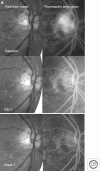
Effect of anti-VEGF agents on retinal neovascularization and macular edema. (A) An intravitreal injection of the anti-VEGF agent bevacizumab has been shown to induce a marked regression of retinal neovascularization beginning as early as 1 day after injection with further regression after 1 week (99). (B) Repeated intravitreal administration of anti-VEGF agents has been shown to reduce the retinal thickening of DME in multiple controlled randomized clinical trials (11,114,116). (A, Derived from Avery et al. 2006; reprinted, with permission, from the author; B, courtesy of the Beetham Eye Institute Image Library, Joslin Diabetes Center, Boston, MA.)
Figure 3.
Figure 3.
Effect of anti-VEGF agents on retinal neovascularization and macular edema. (A) An intravitreal injection of the anti-VEGF agent bevacizumab has been shown to induce a marked regression of retinal neovascularization beginning as early as 1 day after injection with further regression after 1 week (99). (B) Repeated intravitreal administration of anti-VEGF agents has been shown to reduce the retinal thickening of DME in multiple controlled randomized clinical trials (11,114,116). (A, Derived from Avery et al. 2006; reprinted, with permission, from the author; B, courtesy of the Beetham Eye Institute Image Library, Joslin Diabetes Center, Boston, MA.)
Figure 4.
Figure 4.
Among patients with severe NPDR or worse, intravitreal injections of anti-VEGF agents has tended to reduce retinopathy progression and an increase retinopathy regression (Elman et al. 2010). A similar association was also observed in the rates of panretinal photocoagulation and vitreous hemorrhage, which are clinical surrogates of retinopathy progression to proliferative disease (3%, P = 0.002 and 3%, P = 0.02, respectively, vs. 8%). (Figure derived from Elman et al. 2010; reprinted, with permission, from the author.)

References

    1. Abhary S, Burdon KP, Casson RJ, Goggin M, Petrovsky NP, Craig JE 2010. Association between erythropoietin gene polymorphisms and diabetic retinopathy. Arch Ophthalmol 128: 102–106 - PubMed
    1. Abraham P, Yue H, Wilson L 2010. Randomized, double-masked, sham-controlled trial of ranibizumab for neovascular age-related macular degeneration: PIER study year 2. Am J Ophthalmol 150: 315–324 - PubMed
    1. Adamis AP, Aiello LP, D’Amato RA 1999. Angiogenesis and ophthalmic disease. Angiogenesis 3: 9–14 - PubMed
    1. Age-Related Eye Disease Study Research Group 2000. Risk factors associated with age-related macular degeneration. A case-control study in the age-related eye disease study: Age-Related Eye Disease Study Report Number 3. Ophthalmology 107: 2224–2232 - PMC - PubMed
    1. Age-Related Eye Disease Study Research Group 2001. A randomized, placebo-controlled, clinical trial of high-dose supplementation with vitamins C and E, β carotene, and zinc for age-related macular degeneration and vision loss: AREDS report no. 8. Arch Ophthalmol 119: 1417–1436 - PMC - PubMed

MeSH terms

Substances

LinkOut - more resources