. 2012 Jun 7;43(1):50.
doi: 10.1186/1297-9716-43-50.

Improved strategy for phylogenetic analysis of classical swine fever virus based on full-length E2 encoding sequences

Affiliations

Improved strategy for phylogenetic analysis of classical swine fever virus based on full-length E2 encoding sequences

Alexander Postel et al. Vet Res. .

Abstract

Molecular epidemiology has proven to be an essential tool in the control of classical swine fever (CSF) and its use has significantly increased during the past two decades. Phylogenetic analysis is a prerequisite for virus tracing and thus allows implementing more effective control measures. So far, fragments of the 5´NTR (150 nucleotides, nt) and the E2 gene (190 nt) have frequently been used for phylogenetic analyses. The short sequence lengths represent a limiting factor for differentiation of closely related isolates and also for confidence levels of proposed CSFV groups and subgroups. In this study, we used a set of 33 CSFV isolates in order to determine the nucleotide sequences of a 3508-3510 nt region within the 5´ terminal third of the viral genome. Including 22 additional sequences from GenBank database different regions of the genome, comprising the formerly used short 5´NTR and E2 fragments as well as the genomic regions encoding the individual viral proteins Npro, C, Erns, E1, and E2, were compared with respect to variability and suitability for phylogenetic analysis. Full-length E2 encoding sequences (1119 nt) proved to be most suitable for reliable and statistically significant phylogeny and analyses revealed results as good as obtained with the much longer entire 5´NTR-E2 sequences. This strategy is therefore recommended by the EU and OIE Reference Laboratory for CSF as it provides a solid and improved basis for CSFV molecular epidemiology. Finally, the power of this method is illustrated by the phylogenetic analysis of closely related CSFV isolates from a recent outbreak in Lithuania.


Figures

Figure 1
Figure 1
Strategy for RT-PCR and nucleotide sequencing. The 5´-terminal portion of the classical swine fever virus genome encompassing parts of the 5´ nontranslated region (NTR) and the region encoding the N-terminal autoprotease Npro and the structural proteins C, Erns, E1, and E2 was amplified by RT-PCR in three overlapping amplicons (PCR1, PCR2 and PCR3). The analyzed regions include the commonly used 5´NTR fragment (150 nt) and E2 fragment (190 nt) sequences. Location of primers (indicated by arrows) and sequence length of the 5´NTR-E2 region (3508–3510 nt) correspond to the sequence of CSFV strain Alfort187 [GenBank: X87939] and can differ between the isolates due to presence or absence of nucleotides in the 5´NTR. Further information on all primers is given in Table 3.
Figure 2
Figure 2
Ranges of pairwise evolutionary distances among CSFV genotypes, subgenotypes and isolates shown for different genomic regions. Bars represent minimum and maximum values of genetic distances of the entire 5´NTR-E2 region (3508–3510 nt), the full-length E2 encoding sequence (1119 nt), the E2 fragment (190 nt), and the 5´NTR fragment (150 nt). Genetic distances are shown on isolate level (white bars), on subgenotype level (grey bars), and on genotype level (black bars). The analyzed subgenotypes or pairs of subgenotypes or genotypes are indicated by the numbers preceding the bars. With the exception of CSFV “strain 39” [GenBank: AF407339] and strain Great Britain/1964 “Congenital Tremor” [GenBank: JQ411575], which are not assigned to a defined genotype, all sequences shown in the phylogenetic trees of Figure 3 and Figure 4 are included in the analysis. Genetic distances between sequences were calculated by the Kimura-2 parameter method.
Figure 3
Figure 3
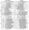
Phylogenetic trees based on the 5´NTR fragment and the entire 5´NTR-E2 sequences. Phylogenetic trees of 33 sequences of isolates from the EURL database (country, year, CSF number) and additional 22 reference sequences originating from GenBank (isolate name, accession number) were calculated by the Neighbor Joining method including bootstrap values for 1000 repetitions. Only statistically significant bootstrap values (≥ 70.0%) are indicated. Evolutionary distances between sequences were calculated by the Kimura-2 parameter method. Trees were rooted at the distinct CSFV strain Great Britain/1964 “Congenital Tremor” [GenBank: JQ411575]. Genotypes and subgenotype names are indicated besides the trees [7,8]. Branch lengths are given as 0.01 substitutions per position according to the scale bars underneath each tree.
Figure 4
Figure 4
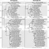
Phylogenetic trees based on the E2 fragment and full-length E2 encoding sequences. Phylogenetic trees of 33 sequences of isolates from the EURL database (country, year, CSF number) and additional 22 reference sequences originating from GenBank (isolate name, accession number) were calculated by the Neighbor Joining method including bootstrap values for 1000 repetitions. Only statistically significant bootstrap values (≥ 70.0%) are indicated. Evolutionary distances between sequences were calculated by the Kimura-2 parameter method. Trees were rooted at the distinct CSFV strain Great Britain/1964 “Congenital Tremor” [GenBank: JQ411575]. Genotypes and subgenotype names are indicated besides the trees [7,8]. Branch lengths are given as 0.01 substitutions per position according to the scale bars underneath each tree.
Figure 5
Figure 5
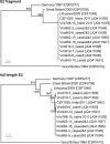
Phylogenetic analysis of recent CSFV isolates from Lithuania. The dendrograms were constructed from the E2 fragment (top) and full-length E2 encoding sequences (bottom) of recent Lithuanian CSFV isolates and selected reference strains of genotype 2.1. Full-length E2 encoding sequences of ten Lithuanian isolates obtained during the outbreak in 2011 were deposited in GenBank [GenBank: JQ411592-JQ411601]. For the Lithuanian isolate obtained in 2009 (CSF1048, Panevezys”) the 5´NTR-E2 sequence was determined [GenBank: JQ411591] and found to be identical to a previously published full-genome sequence [GenBank: HQ148063]. Trees were rooted at strain Great Britain/1964 “Congenital Tremor” [GenBank: JQ411575]. Distances were calculated by the Kimura-2 parameter method and used to construct the trees according to the Neighbor joining method. Trees are drawn to scale as indicated by the respective scale bars (0.01 substitutions per position). Bootstrap values were generated by 1000 repetitions; only statistically significant values (≥ 70.0%) are indicated.

Similar articles

Cited by

References

    1. Edwards S, Fukusho A, Lefevre PC, Lipowski A, Pejsak Z, Roehe P, Westergaard J. Classical swine fever: the global situation. Vet Microbiol. 2000;73:103–119. doi: 10.1016/S0378-1135(00)00138-3. - DOI - PubMed
    1. Depner KR, Strebelow G, Staubach C, Kramer M, Teuffert J, Botcher L, Hoffmann B, Beer M, Greiser-Wilke I, Mettenleiter T. Case report: the significance of genotyping for the epidemiological tracing of classical swine fever (CSF) Dtsch Tierarztl Wochenschr. 2006;113:159–162. - PubMed
    1. Moennig V, Floegel-Niesmann G, Greiser-Wilke I. Clinical signs and epidemiology of classical swine fever: a review of new knowledge. Vet J. 2003;165:11–20. doi: 10.1016/S1090-0233(02)00112-0. - DOI - PubMed
    1. Hoffmann B, Beer M, Schelp C, Schirrmeier H, Depner K. Validation of a real-time RT-PCR assay for sensitive and specific detection of classical swine fever. J Virol Methods. 2005;130:36–44. doi: 10.1016/j.jviromet.2005.05.030. - DOI - PubMed
    1. Vilcek S, Herring AJ, Herring JA, Nettleton PF, Lowings JP, Paton DJ. Pestiviruses isolated from pigs, cattle and sheep can be allocated into at least three genogroups using polymerase chain reaction and restriction endonuclease analysis. Arch Virol. 1994;136:309–323. doi: 10.1007/BF01321060. - DOI - PubMed

Publication types

Associated data