. 2012 Jun 7:7:34.
doi: 10.1186/1750-1172-7-34.

Novel genetic linkage of rat Sp6 mutation to Amelogenesis imperfecta

Affiliations

Novel genetic linkage of rat Sp6 mutation to Amelogenesis imperfecta

Taro Muto et al. Orphanet J Rare Dis. .

Abstract

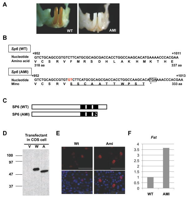
Background: Amelogenesis imperfecta (AI) is an inherited disorder characterized by abnormal formation of tooth enamel. Although several genes responsible for AI have been reported, not all causative genes for human AI have been identified to date. AMI rat has been reported as an autosomal recessive mutant with hypoplastic AI isolated from a colony of stroke-prone spontaneously hypertensive rat strain, but the causative gene has not yet been clarified. Through a genetic screen, we identified the causative gene of autosomal recessive AI in AMI and analyzed its role in amelogenesis.

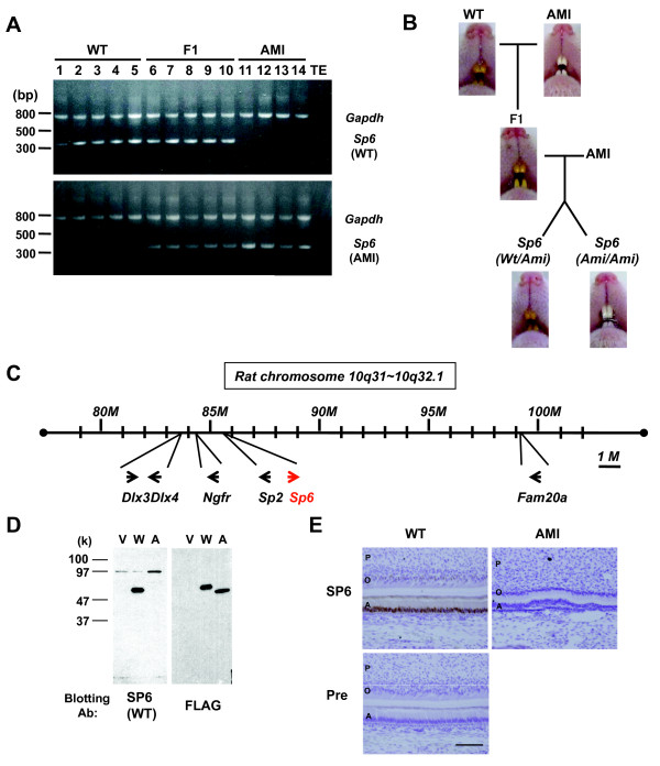
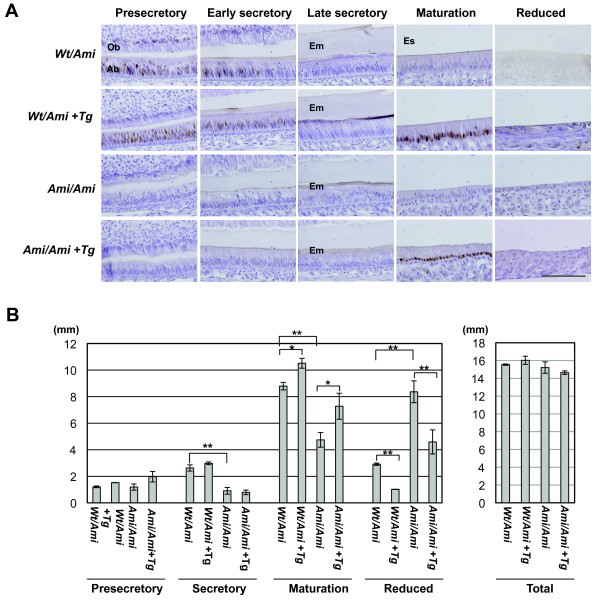
Methods: cDNA sequencing of possible AI-candidate genes so far identified using total RNA of day 6 AMI rat molars identified a novel responsible mutation in specificity protein 6 (Sp6). Genetic linkage analysis was performed between Sp6 and AI phenotype in AMI. To understand a role of SP6 in AI, we generated the transgenic rats harboring Sp6 transgene in AMI (Ami/Ami + Tg). Histological analyses were performed using the thin sections of control rats, AMI, and Ami/Ami + Tg incisors in maxillae, respectively.

Results: We found the novel genetic linkage between a 2-bp insertional mutation of Sp6 gene and the AI phenotype in AMI rats. The position of mutation was located in the coding region of Sp6, which caused frameshift mutation and disruption of the third zinc finger domain of SP6 with 11 cryptic amino acid residues and a stop codon. Transfection studies showed that the mutant protein can be translated and localized in the nucleus in the same manner as the wild-type SP6 protein. When we introduced the CMV promoter-driven wild-type Sp6 transgene into AMI rats, the SP6 protein was ectopically expressed in the maturation stage of ameloblasts associated with the extended maturation stage and the shortened reduced stage without any other phenotypical changes.

Conclusion: We propose the addition of Sp6 mutation as a new molecular diagnostic criterion for the autosomal recessive AI patients. Our findings expand the spectrum of genetic causes of autosomal recessive AI and sheds light on the molecular diagnosis for the classification of AI. Furthermore, tight regulation of the temporospatial expression of SP6 may have critical roles in completing amelogenesis.


Figures

Figure 1
Figure 1
Identification of a 2-bp insertion inSp6 in AMI rats. A. Representative phenotype of WT and AMI rats. Photographs show mandibular incisors from each of the rats. B. The nucleotide sequence of Sp6 in wild-type (WT) and AMI rats. The 2-bp insertion is highlighted (red). The square represents the premature stop codon. The amino acid sequence deduced from the codons is shown below the nucleotide sequence. The altered amino acid sequence in Ami-SP6 is underlined. C. Schematic representation of SP6. The N-terminal region is located to the left. Black boxes represent the position of the zinc finger motifs. The box with diagonal lines shows the altered amino acid sequence in Ami-SP6. D. Western blot analyses. COS7 cells were transfected with an expression vector only (V) or with FLAG-tagged Wt- (W) or Ami- (A) cDNA. Samples were blotted with FLAG antibodies. E. Subcellular localization of FLAG-tagged SP6 by immunocytochemical analysis. Red, FLAG-tagged protein; Blue, Nuclei. F. Relative expression levels of Fst in WT and AMI molars.
Figure 2
Figure 2
Linkage analysis betweenSp6 and AI in AMI rats. A. Genomic PCR products amplified by WT- (upper panel) or AMI- (lower panel) Sp6-specific primers. Sample numbers and the characteristics of the animals are denoted above the gel image. B. The color of the incisors was examined. Pups derived from the cross between F1 and AMI rats were sorted by the Sp6 genotype to determine the correlation between Sp6 and AI in AMI rats (Wt/Ami, n = 62; Ami/Ami, n = 56). C. Schematic diagram of focal gene localization in rat chromosome 10q31–10q32.1. D. Western blot analyses. COS7 cells were transfected with expression vector only (V) or with FLAG-tagged Wt- (W) or Ami- (A) cDNA. Samples were blotted with the indicated antibodies. E. Immunohistochemical analysis of incisors from newborn pups. Sections from secretory stage incisors of WT rats and those of the corresponding region from AMI rats were stained with the anti-Wt-SP6 antiserum (SP6) or preimmune rabbit serum (Pre). Signals were obtained from DAB (brown). Sections were counterstained with Hematoxylin. Scale bar: 100 mm. A, ameloblasts; O, odontoblasts; P, pulp.
Figure 3
Figure 3
Analyses of longitudinal sections of the maxillary incisors of 6-week-old rats. A. Immunohistochemical analysis. Incisor sections were immunostained with antiserum against rat Wt-SP6. The sections were prepared from rats heterozygous (Wt/Ami) or homozygous (Ami/Ami) for mutant Sp6. Animals were sorted based on the transgenic Sp6 (Tg) genotype. Scale bar, 100 mm. Ab, ameloblasts; Em, enamel matrix; Es, enamel space; Ob, odontoblasts. B. The length of the ameloblast layer for the indicated differentiation stages. Columns represent the average of two to three independent samples; bars indicate standard deviation. Statistical significance was evaluated by unpaired t tests for an indicated set of data. *p < 0.05, **p < 0.01.
Figure 4
Figure 4
Organogenesis requiringSp6 activity. A. Whiskers of rats at postnatal day 1. Each Sp6 genotype in the 1st backcrossed generation was examined. (Wt/Ami, n = 13; Ami/Ami, n = 21) B. Whiskers of adult rats. WT and AMI rats were examined when they were 6–8 months old (WT, n = 3; AMI, n = 14) C. X-ray analysis of the maxillae (Max) and mandibles (Man) of WT (1, 2) and AMI (3, 4) rats. Molars (1, 3) and incisors (2, 4) are highlighted (WT, n = 2; AMI, n = 2) D. Normal number of digits in AMI rats. Digits of the left (1, 3) and right (2, 4) limbs of WT (1, 2) and AMI (3, 4) rats are shown. Upper panels, forelimb; lower panels, hindlimb (WT, n = 3; AMI, n = 14).

Similar articles

Cited by

References

    1. Crawford PJ, Aldred M, Bloch-Zupan A. Amelogenesis imperfecta. Orphanet J Rare Dis. 2007;2:17. - PMC - PubMed
    1. Witkop CJ Jr. Amelogenesis imperfecta, dentinogenesis imperfecta and dentin dysplasia revisited: problems in classification. J Oral Pathol. 1988;17:547–553. - PubMed
    1. Cho SH, Seymen F, Lee KE, Lee SK, Kweon YS, Kim KJ, Jung SE, Song SJ, Yildirim M, Bayram M, Tuna EB, Gencay K, Kim JW. Novel FAM20A mutations in hypoplastic amelogenesis imperfecta. Hum Mutat. 2011;33:91–94. - PubMed
    1. O'Sullivan J, Bitu CC, Daly SB, Urquhart JE, Barron MJ, Bhaskar SS, Martelli-Junior H, dos Santos Neto PE, Mansilla MA, Murray JC, Coletta RD, Black GC, Dixon MJ. Whole-Exome sequencing identifies FAM20A mutations as a cause of amelogenesis imperfecta and gingival hyperplasia syndrome. Am J Hum Genet. 2011;88:616–620. - PMC - PubMed
    1. Santos MC, Hart PS, Ramaswami M, Kanno CM, Hart TC, Line SR. Exclusion of known gene for enamel development in two Brazilian families with amelogenesis imperfecta. Head Face Med. 2007;3:8. - PMC - PubMed

Publication types

LinkOut - more resources