. 2012 Sep 20;29(14):2375-92.
doi: 10.1089/neu.2012.2408. Epub 2012 Jul 12.

A CD11d monoclonal antibody treatment reduces tissue injury and improves neurological outcome after fluid percussion brain injury in rats

Affiliations

A CD11d monoclonal antibody treatment reduces tissue injury and improves neurological outcome after fluid percussion brain injury in rats

Feng Bao et al. J Neurotrauma. .

Abstract

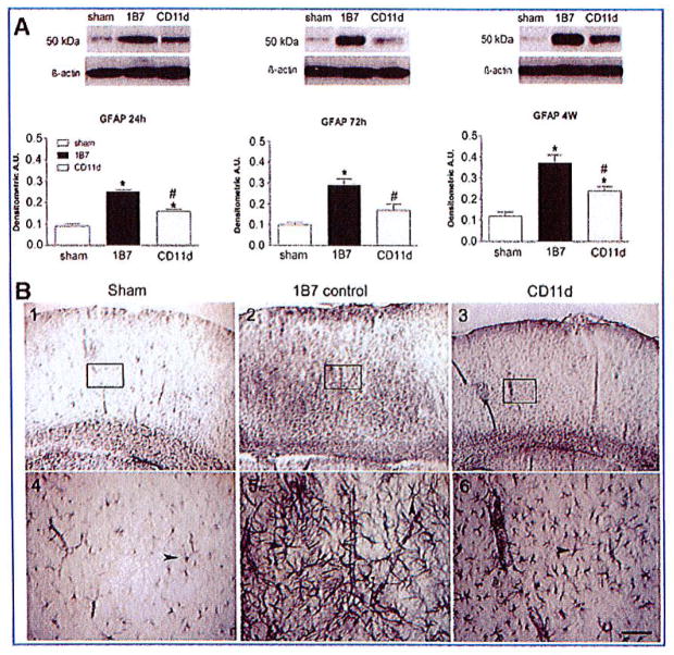
Traumatic brain injury (TBI) is an international health concern often resulting in chronic neurological abnormalities, including cognitive deficits, emotional disturbances, and motor impairments. An anti-CD11d monoclonal antibody that blocks the CD11d/CD18 integrin and vascular cell adhesion molecule (VCAM)-1 interaction following experimental spinal cord injury improves functional recovery, while reducing the intraspinal number of neutrophils and macrophages, oxidative activity, and tissue damage. Since the mechanisms of secondary injury in the brain and spinal cord are similar, we designed a study to evaluate fully the effects of anti-CD11d treatment after a moderate lateral fluid percussion TBI in the rat. Rats were treated at 2 h after TBI with either the anti-CD11d antibody or an isotype-matched control antibody 1B7, and both short (24- to 72-h) and long (4-week) recovery periods were examined. The anti-CD11d integrin treatment reduced neutrophil and macrophage levels in the injured brain, with concomitant reductions in lipid peroxidation, astrocyte activation, amyloid precursor protein accumulation, and neuronal loss. The reduced neuroinflammation seen in anti-CD11d-treated rats correlated with improved performance on a number of behavioral tests. At 24 h, the anti-CD11d group performed significantly better than the 1B7 controls on several water maze measures of spatial cognition. At 4 weeks post-injury the anti-CD11d-treated rats had better sensorimotor function as assessed by the beam task, and reduced anxiety-like behaviors, as evidenced by elevated-plus maze testing, compared to 1B7 controls. These findings suggest that neuroinflammation is associated with behavioral deficits after TBI, and that anti-CD11d antibody treatment is a viable strategy to improve neurological outcomes after TBI.


Figures

FIG. 1
FIG. 1
Histological sections of injured brain stained with hematoxylin and eosin 24 h after fluid percussion injury (FPI). The section is at the location of the FPI and includes the hippocampus. Areas boxed in low-power photomicrographs (A–C) are shown at high-power in panels D–F. Sections from the 1B7 control rat and the anti-CD11d-treated rat show areas of tissue disruption and hemorrhage immediately above and including the hippocampus (arrowheads; scale bar in A–C = 200 μm; scale bar in D–F = 100 μm).
FIG. 2
FIG. 2
Neutrophil infiltration into the injured brain is reduced by the anti-CD11d treatment. (A) Photomicrographs of the cortex centered on the lesion epicenter (or a comparable position in sham-injured rats), immunostained by an anti-neutrophil antibody. The sections from sham-injured, 1B7 control, and anti-CD11d-treated rats are shown at low power in panels 1–3, respectively, and the boxed areas are shown at high power in panels 4–6 (scale bar in A1–3 = 100 μm, and in A4–6 = 100 μm). (B) Left panel shows counts of neutrophils within a 0.2-mm2 area of interest centered on the epicenter in the injured brain in the sham, 1B7 control, and anti-CD11d-treated groups (n = 4/group) at 24 h after injury (*significantly different from sham animals, and #significantly different from the 1B7 control group, p ≤ 0.05 after the Student Neuman Keuls test and one-way analysis of variance [ANOVA]; ANOVA for neutrophil count: F2,9 = 8.6, p = 0.008). (B) Right panels show myeloperoxidase (MPO) activity in brain homogenates after traumatic brain injury (TBI) at 24 h in sham-injured animals (n = 6), 1B7 controls, and anti-CD11d animals (n = 7/group); at 72 h in these groups (n = 7, 8, and 7, respectively); and at 4 weeks (n = 5, 6, and 5, respectively; ANOVA: 24 h, F2,17 = 13.63, p < 0.001; 72 h, F2,19 = 11.75, p < 0.001; 4 weeks, F2,13 = 2.74, p = 0.102). (C) Neutrophil protein identified by Western blotting in brain homogenates expressed as means ± standard error with a representative autoradiogram shown above each set of histograms. Neutrophil protein is shown at 24 h, 72 h, and 4 weeks in sham-injured, 1B7 controls, and anti-CD11d groups (n = 5/group; ANOVA: 24 h, F2,12 = 20.52, p < 0.001; 72 h, F2,12 = 20.70, p < 0.001; 4 weeks, F2,12 = 2.92, p = 0.09; A.U., arbitrary units). Color image is available online at www.liebertonline.com/neu
FIG. 3
FIG. 3
Macrophages in the injured brain are reduced by the anti-CD11d treatment. (A and B) Photomicrographs 72 h or 4 weeks post-injury of the cortex centered on the lesion epicenter (or a comparable position in sham-injured rats), immunostained by an anti-ED-1 antibody. The sections from sham-injured, 1B7 control, and anti-CD11d-treated rats are shown at low power in panels 1–3, respectively, and the boxed areas are shown at high power in panels 4–6 (*significantly different from sham animals, and #significantly different from the 1B7 control group, p ≤ 0.05 after the Student Neuman Keuls test and one-way analysis of variance; scale bar in A and B1–3 = 100 μm, and in A and B4–6 = 100 μm). Arrows indicate positions of blood vessels, and the arrowhead indicates a typical ED-1-expressing macrophage. (C) Left panel shows counts of macrophages in a 0.2-mm2 area within the injured brain in the sham, 1B7 control, and anti-CD11d-treated groups at 72 h (n = 4/group; analysis of variance [ANOVA]: F2,9 = 61.34, p < 0.001). Right panel shows counts of macrophages at 4 weeks after traumatic brain injury (TBI) (n = 7, 6, and 6, respectively; ANOVA: F2,16 = 7.04, p = 0.006). (D) Macrophage protein identified by Western blotting in cord homogenates expressed as mean values ± standard error, with a representative autoradiogram shown above each set of histograms. Macrophage protein is shown at 24 h, 72 h, and 4 weeks in the sham-injured, 1B7 control, and anti-CD11d groups (n = 5/group; ANOVA: 24 h, F2,12 = 7.15, p = 0.009; 72 h, F2,12 = 17.44, p < 0.001; 4 weeks, F2,12 = 15.95, p < 0.001; A.U., arbitrary units). Color image is available online at www.liebertonline.com/neu
FIG. 4
FIG. 4
Lipid peroxidation is reduced by the anti-CD11d treatment. TBARS concentrations at 24 h after TBI in the sham-injured (n = 6), 1B7 control, and anti-CD11d (n = 7 each) groups; at 72 h in these groups (n = 7, 7, and 8, respectively), and at 4 weeks (n = 5, 6, and 5, respectively; analysis of variance [ANOVA]: 24 h, F2,17 = 4.68, p = 0.024; 72 h, F2,19 = 5.10, p = 0.017; 4 weeks, F2,13 = 5.3, p = 0.021; ANOVA, analysis of variance; TBI, traumatic brain injury; TBARS, thiobarbituric acid reactive substances; *significantly different from sham animals, and #significantly different from the 1B7 control group, p ≤ 0.05 after the Student Neuman Keuls test and one-way ANOVA).
FIG. 5
FIG. 5
Astrogliosis assessed by the expression of glial fibrillary acidic protein (GFAP) in the injured brain is reduced by the anti-CD11d treatment. (A) GFAP, identified by Western blotting in cord homogenates, is shown at 24 h, 72 h, and 4 weeks, in the sham-injured, 1B7 control, and anti-CD11d groups (n = 5/group; analysis of variance [ANOVA]: 24 h, F2,12 = 35.75, p < 0.001; 72 h, F2,12 = 15.38, p < 0.001; 4 weeks, F2,12 = 22.27, p < 0.001). (B) Shown are photomicrographs of brain sections at the location of the fluid percussion injury immunostained by an antibody to GFAP. Boxed areas in panels 1–3 are shown at higher power in panels 4–6 (scale bars = 100 μm in A1–3 and in A4–6). Arrowheads indicate examples of reactive astrocytes. Note the increased intensity of anti-GFAP staining and the presence of overlapping, hypertrophic GFAP-positive processes in the sections from 1B7 controls (panels 2 and 5), compared to anti-CD11d treated rats (panels 3 and 6; *significantly different from sham animals, and #significantly different from the 1B7 control group, p ≤ 0.05 after the Student Neuman Keuls test and one-way ANOVA; A.U., arbitrary units). Color image is available online at www.liebertonline.com/neu
FIG. 6
FIG. 6
The increase in amyloid precursor protein (APP) expression in the injured brain is reduced by the anti-CD11d treatment. (A) APP, identified by Western blotting in brain homogenates, is shown at 24 h, 72 h, and 4 weeks, in the sham-injured, 1B7 control, and anti-CD11d groups (n = 5/group; analysis of variance [ANOVA]: 24 h, F2,12 = 10.46, p = 0.002; 72 h, F2,12 = 22.11, p < 0.001; 4 weeks, F2,12 = 9.04, p = 0.004). (B) Photomicrographs of the cortex centered on the lesion epicenter (or a comparable position in sham-injured rats) immunostained by an antibody to APP. Boxed areas in panels 1–3 are shown at high power in panels 4–6 (scale bars = 100 μm in A1–3 and A4–6). Arrowheads indicate APP-positive aggregates (*significantly different from sham animals, and #significantly different from the 1B7 control group, p ≤ 0.05 after the Student Neuman Keuls test and one-way ANOVA; A.U., arbitrary units). Color image is available online at www.liebertonline.com/neu
FIG. 7
FIG. 7
Expression of neuronal nuclear antigen (NeuN) in the injured brain. NeuN, identified by Western blotting in brain homogenates, is shown at 24 h, 72 h, and 4 weeks in the sham-injured, 1B7 control, and anti-CD11d groups (n = 5/group; analysis of variance [ANOVA]: 24 h, F2,12 = 10.42, p = 0.002; 72 h, F2,12 = 28.96, p < 0.001; 4 weeks, F2,12 = 12.52, p = 0.001; *significantly different from sham animals, and #significantly different from the 1B7 control group, p ≤ 0.05 after the Student Neuman Keuls test and one-way ANOVA; A.U., arbitrary units).
FIG. 8
FIG. 8
Counts of neurons in the hippocampus identified by neuronal nuclear antigen (NeuN). (A) Photomicrographs of sections of the hippocampus within the region of the FPI immunostained by an antibody to NeuN. The sections from the sham-injured, 1B7 control, and anti-CD11d groups are shown at low power in panels 1–3, and boxed areas are shown at high power in panels 4–6 (scale bars = 100 μm in A1-3 and A4-6). (B) Counts of neurons in a 0.33-mm2 area of interest (boxed areas in the images) within the hippocampus. Neuron counts are shown at 24h in the sham-injured, 1B7 control, and anti-CD11d groups (n = 4/group) at 24h, 72h (n = 5/group), and 4 weeks (n=9, 8, and 7, respectively; analysis of variance [ANOVA]: 24h, F2,9 = 0.06, p = 0.943; 72h, F2,12 = 0.15, p = 0.865; 4 weeks, F2,21=4.35, p = 0.026; *Significantly different from sham animals, and #significantly different from the 1B7 control group, p≤0.05 after the Student Neuman Keuls test and one-way ANVOVA). Color image is available online at www.liebertonline.com/neu
FIG. 9
FIG. 9
The anti-CD11d treatment improved spatial cognition in the short recovery (SR) group as assessed by water maze testing. (A) Graph of search times for the acquisition task in the SR analysis for sham-injured rats (SHAM-SR, open triangles), 1B7-treated rats (1B7-SR, gray squares), and anti-CD11d-treated rats (CD11d-SR, solid black circles). Each block plotted on the x-axis represents the average of results of 2 of the 10 trials (n = 12 rats/group; *anti-CD11d-SR and 1B7-SR groups had significantly longer search times than the sham-SR group). (B) Percent of direct and circle swims in the acquisition task in the SR analysis for the three groups of rats. Histogram bars represent means of data during 10 water maze trials (*1B7-SR rats had significantly fewer direct and circle swims than the sham-SR rats; analysis of variance [ANOVA]: F2,36 = 5.616, p<0.01). (C) Swim speeds during these trials of acquisition testing did not differ between the three groups. (D) Search times for the reversal task in the SR analysis of reversal training (format and number of rats as in A; #the anti CD11d-SR group had significantly shorter search times compared to the 1B7-SR group, but did not differ from the sham-SR group; *the 1B7-SR group had significantly longer search times than the sham-SR group). (E) Direct and circle swims in the reversal task for the three groups of SR rats (#significantly more direct and circle swims than the 1B7-SR group; *different from sham-SR; ANOVA: F2,36 = 4.223, p<0.05). The anti-CD11d-SR group did not differ from the sham-SR group. (F) Swim speeds during these trials of acquisition training did not differ between the three groups.
FIG. 10
FIG. 10
Anti-CD11d treatment improved spatial cognition in the long recovery (LR) water maze. (A and D) No differences in search time in the acquisition or reversal tasks were found among the sham-injured rats (SHAM-SR, open triangles), 1B7-treated rats (1B7-SR, gray squares), and anti-CD11d-treated rats (CD11d-SR, solid black circles; n = 7 rats/group) tested 4 weeks post-injury. (B and E) Percentages of direct and circle swims in the study groups during acquisition and reversal training. Histogram bars represent means of data collected during the 10 water maze trials (*significantly different from sham-LR rats; F2,41 =4.346, p<0.05). The CD11d-LR group was no different from the sham-LR group. (C and F) Swim speeds during acquisition and reversal training were no different between the groups.
FIG. 11
FIG. 11
The anti-CD11d treatment improved long recovery (LR) beam task performance. (A) Number of slips and falls during the beam task in the sham-short recovery (SHAM-SR), 1B7-SR, and CD11d-SR groups of rats (n = 12/group). Histogram bars represent means of data collected during the 10 beam task trials (*significantly different from the sham-SR group; F2,36=3.748, p<0.05). The CD11d-SR group did not differ from sham-SR group. (B) Number of slips and falls in the LR groups (n =7/group; #significantly different from the 1B7-LR group; *different from the sham-LR group; F2,41 = 4.526, p<0.05). The CD11d-LR group did not differ from the sham-LR group.
FIG. 12
FIG. 12
Anti-CD11d treatment reduces anxiety in long recovery (LR) testing. Time spent in the open arm of the maze (reciprocally related to anxiety; A), and number of entries into the closed arm (B), by the short recovery (SR) sham-injured (SHAM-SR, n =24), 1B7 (1B7-SR, n = 24), and anti-CD11d (CD11d-SR, n = 25) groups of rats. Histogram bars represent time or entries accrued during 5 min of free exploration. No significant differences were detected among the groups at this time. Time spent in the open arm of the maze (C), and number of closed arm entries (D), by the LR groups of rats (n = 14 rats/group; #significantly different from the 1B7-LR group; *significantly different from the sham-LR group; F2,41 =3.925, p<0.05). The CD11d-LR group did not differ from the sham-LR group in time spent in the open arm. The three LR groups did not differ from each other in the number of entries into the closed arm (SHAM-LR, sham-long recovery; 1B7-LR, 1B7-long recovery; CD11d-LR, CD11d-long recovery).

Similar articles

Cited by

References

    1. Aiguo W, Zhe Y, Gomez-Pinilla F. Vitamin E protects against oxidative damage and learning disability after mild traumatic brain injury in rats. Neurorehabil Neural Repair. 2010;24:290–298. - PMC - PubMed
    1. Atif F, Sayeed I, Ishrat T, Stein DG. Progesterone with vitamin D affords better neuroprotection against excitotoxicity in cultured cortical neurons than progesterone alone. Molecular Med. 2009;15:328–336. - PMC - PubMed
    1. Bao F, Chen Y, Dekaban GA, Weaver LC. An anti-CD11d integrin antibody reduces cyclooxygenase-2 expression and protein and DNA oxidation after spinal cord injury in rats. J Neurochem. 2004a;90:1194–1204. - PubMed
    1. Bao F, Chen Y, Dekaban GA, Weaver LC. Early anti-inflammatory treatment reduces lipid peroxidation and protein nitration after spinal cord injury in rats. J Neurochem. 2004b;88:1335–1344. - PubMed
    1. Bao F, Dekaban GA, Weaver LC. Anti-CD11d antibody treatment reduces free radical formation and cell death in the injured spinal cord of rats. J Neurochem. 2005;94:1361–1373. - PubMed

This article has been cited by

    1. Zhang B, Gensel JC. Is neuroinflammation in the injured spinal cord different than in the brain? Examining intrinsic differences between the brain and spinal cord. Experimental Neurology. 2014;258:112–120. - PubMed
    1. Shultz Sandy R, TanXin L, Wright David K, Liu Shijie J, Semple Bridgette D, Leigh Johnston, Jones Nigel C, Cook Andrew D, Hamilton John A, O’Brien Terence J. Granulocyte-Macrophage Colony-Stimulating Factor Is Neuroprotective in Experimental Traumatic Brain Injury. Journal of Neurotrauma. 2014;31:10. - PMC - PubMed
    1. John C. GenselSpinal Cord Injury. :339–361.
    1. Gold Eric M, Su Diane, López-Velázquez Luci, Haus Daniel L, Perez Harvey, Lacuesta George A, Anderson Aileen J, Cummings Brian J. Functional assessment of long-term deficits in rodent models of traumatic brain injury. Regenerative Medicine. 2013;8:4. - PubMed
    1. Shultz Sandy R, Bao Feng, Weaver Lynne C, Cain Donald P, Brown Arthur. Treatment with an anti-CD11d integrin antibody reduces neuroinflammation and improves outcome in a rat model of repeated concussion. Journal of Neuroinflammation. 2013;10:1. - PMC - PubMed

Publication types

Grants and funding

LinkOut - more resources