

Ultra-high resolution diffusion tensor imaging of the microscopic pathways of the medial temporal lobe
- PMID: 22677150
- PMCID: PMC4315156
- DOI: 10.1016/j.neuroimage.2012.05.065
Ultra-high resolution diffusion tensor imaging of the microscopic pathways of the medial temporal lobe
Abstract
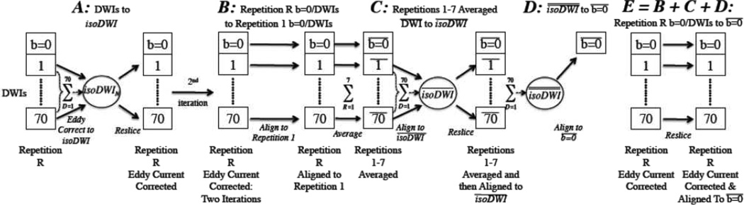
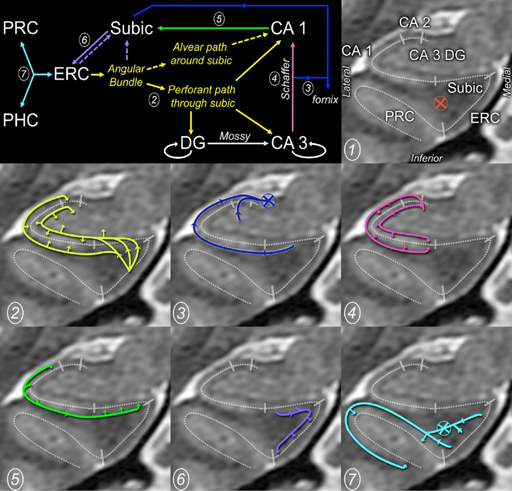
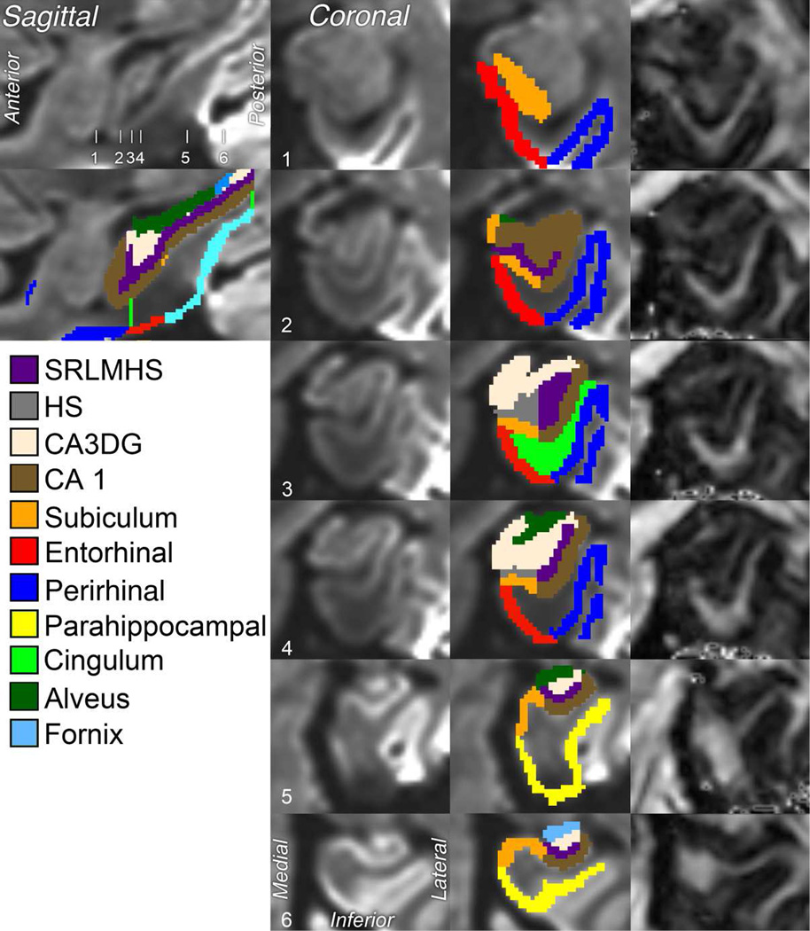
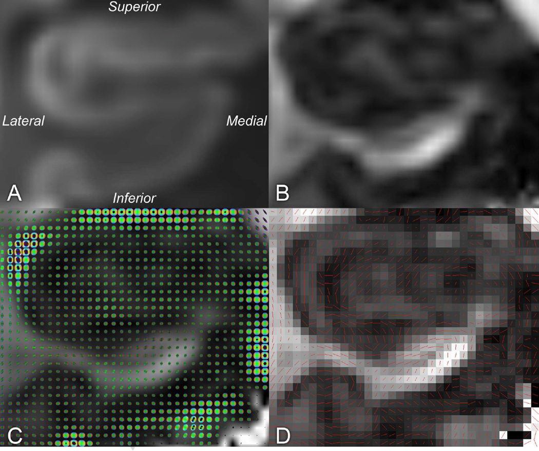
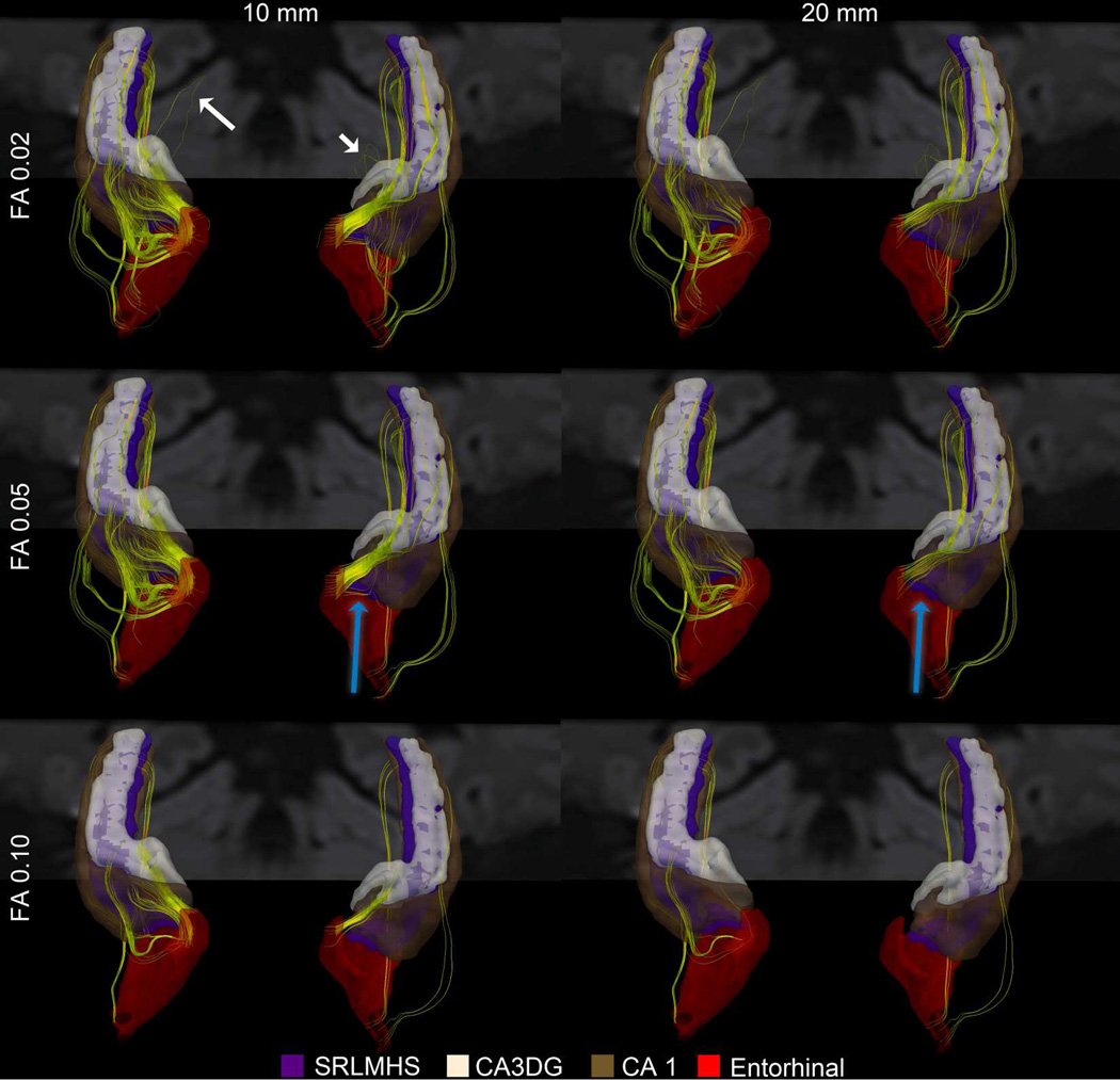
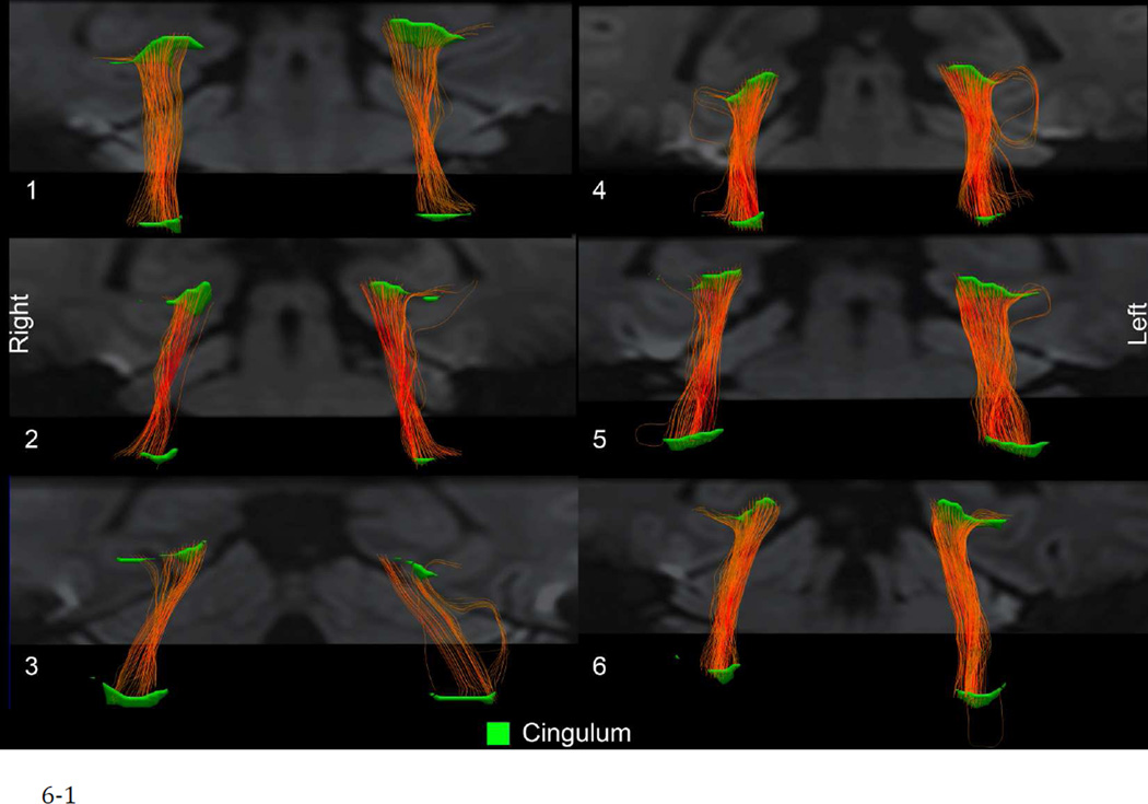
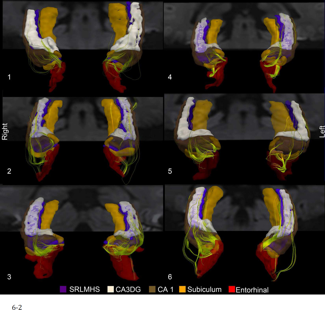
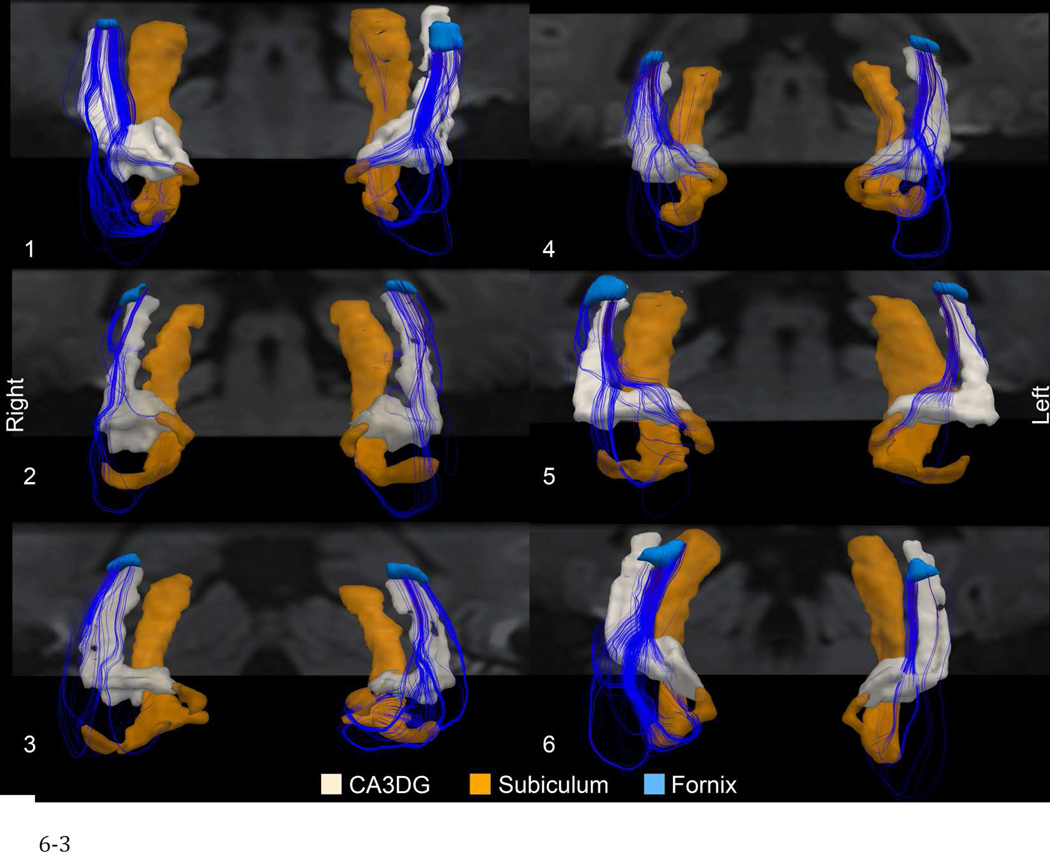
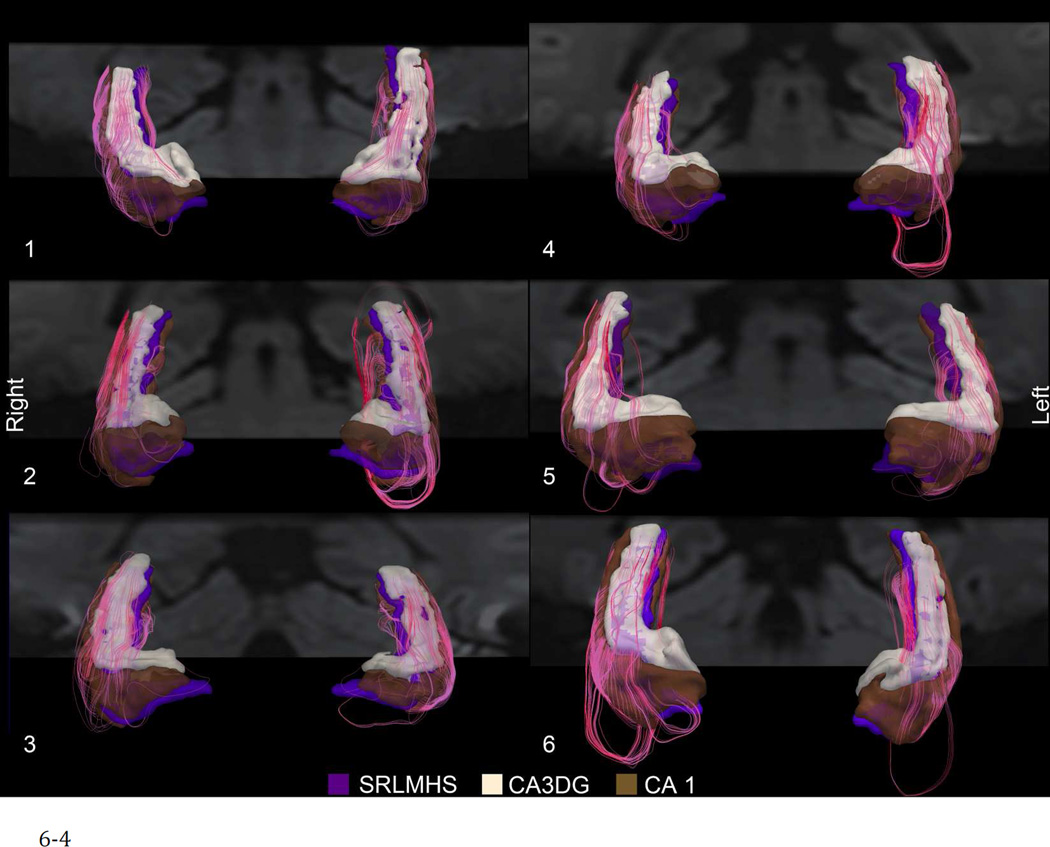
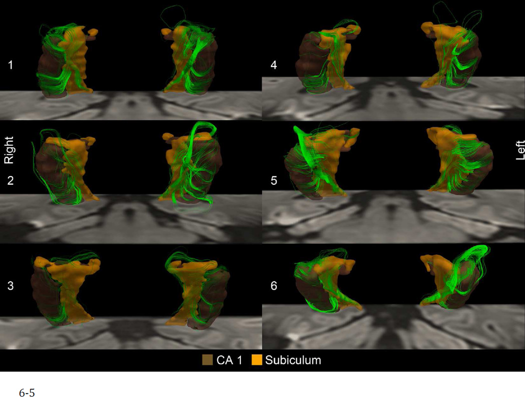
Diseases involving the medial temporal lobes (MTL) such as Alzheimer's disease and mesial temporal sclerosis pose an ongoing diagnostic challenge because of the difficulty in identifying conclusive imaging features, particularly in pre-clinical states. Abnormal neuronal connectivity may be present in the circuitry of the MTL, but current techniques cannot reliably detect those abnormalities. Diffusion tensor imaging (DTI) has shown promise in defining putative abnormalities in connectivity, but DTI studies of the MTL performed to date have shown neither dramatic nor consistent differences across patient populations. Conventional DTI methodology provides an inadequate depiction of the complex microanatomy present in the medial temporal lobe because of a typically employed low isotropic resolution of 2.0-2.5 mm, a low signal-to-noise ratio (SNR), and echo-planar imaging (EPI) geometric distortions that are exacerbated by the inhomogeneous magnetic environment at the skull base. In this study, we pushed the resolving power of DTI to near-mm isotropic voxel size to achieve a detailed depiction of mesial temporal microstructure at 3 T. High image fidelity and SNR at this resolution are achieved through several mechanisms: (1) acquiring multiple repetitions of the minimum field of view required for hippocampal coverage to boost SNR; (2) utilizing a single-refocused diffusion preparation to enhance SNR further; (3) performing a phase correction to reduce Rician noise; (4) minimizing distortion and maintaining left-right distortion symmetry with axial-plane parallel imaging; and (5) retaining anatomical and quantitative accuracy through the use of motion correction coupled with a higher-order eddy-current correction scheme. We combined this high-resolution methodology with a detailed segmentation of the MTL to identify tracks in all subjects that may represent the major pathways of the MTL, including the perforant pathway. Tractography performed on a subset of the data identified similar tracks, although they were lesser in number. This detailed analysis of MTL substructure may have applications to clinical populations.
Copyright © 2012 Elsevier Inc. All rights reserved.
Figures
References
-
- Aggleton JP, Brown MW. Episodic memory, amnesia, and the hippocampal-anterior thalamic axis. Behav Brain Sci. 1999;22:425–444. discussion 444–489. - PubMed
-
- Alexander DC, Barker GJ. Optimal imaging parameters for fiber-orientation estimation in diffusion MRI. Neuroimage. 2005;27:357–367. - PubMed
-
- Amaral DG, Dolorfo C, Alvarez-Royo P. Organization of CA1 projections to the subiculum: a PHA-L analysis in the rat. Hippocampus. 1991;1:415–435. - PubMed
-
- Andersson JLR, Skare S. A model-based method for retrospective correction of geometric distortions in diffusion-weighted EPI. Neuroimage. 2002;16:177–199. - PubMed
Publication types
MeSH terms
Grants and funding
LinkOut - more resources
Full Text Sources
