. 2012 May 16;102(10):2401-10.
doi: 10.1016/j.bpj.2012.03.065. Epub 2012 May 15.

Correlating calcium binding, Förster resonance energy transfer, and conformational change in the biosensor TN-XXL

Affiliations

Correlating calcium binding, Förster resonance energy transfer, and conformational change in the biosensor TN-XXL

Anselm Geiger et al. Biophys J. .

Abstract

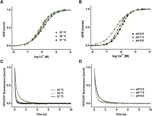
Genetically encoded calcium indicators have become instrumental in imaging signaling in complex tissues and neuronal circuits in vivo. Despite their importance, structure-function relationships of these sensors often remain largely uncharacterized due to their artificial and multimodular composition. Here, we describe a combination of protein engineering and kinetic, spectroscopic, and biophysical analysis of the Förster resonance energy transfer (FRET)-based calcium biosensor TN-XXL. Using fluorescence spectroscopy of engineered tyrosines, we show that two of the four calcium binding EF-hands dominate the FRET output of TN-XXL and that local conformational changes of these hands match the kinetics of FRET change. Using small-angle x-ray scattering and NMR spectroscopy, we show that TN-XXL changes from a flexible elongated to a rigid globular shape upon binding calcium, thus resulting in FRET signal output. Furthermore, we compare calcium titrations using fluorescence lifetime spectroscopy with the ratiometric approach and investigate potential non-FRET effects that may affect the fluorophores. Thus, our data characterize the biophysics of TN-XXL in detail and may form a basis for further rational engineering of FRET-based biosensors.


Figures

Figure 1
Figure 1
Fluorescence spectroscopy of single tyrosine substitutions in the calcium binding domain of TN-XXL. (A) Schematic representation of the four calcium binding EF-hands of the TN-XXL calcium binding domain. Endogenous phenylalanine residues were mutated to tyrosines to report local calcium binding at individual EF-hands. (B) Calcium titrations of single tyrosine substitutions in comparison to TN-XXL FRET. Standard error of the mean depicted as error bars and fluorescence normalized. (C) Calcium-dissociation kinetics of EF3-1 and EF3-2. Tyrosine measurements used excitation at 275 nm with emission recorded at 303 nm. TN-XXL FRET control was performed with excitation at 432 nm and cpCitrine (YFP) emission was recorded at 527 nm. All data are averages of three independent experiments.
Figure 2
Figure 2
Hydrodynamics of TN-XXL. (A) Coomassie staining of SDS- and native PAGE gels of purified TN-XXL. (B) Size-exclusion chromatography of TN-XXL in Ca2+-free and high-Ca2+ conditions on a Superose 12 column (10/300). (C) c(S) distribution calculated using SEDFIT (28) from sedimentation-velocity experiments in the analytical ultracentrifuge with TN-XXL concentrations of 18 and 23 μM in the presence and absence, respectively, of Ca2+. (D) Solution scattering data for TN-XXL in Ca2+-free and high-Ca2+ conditions. (E and F) Distance distribution functions P(r) and Kratky plot, respectively, of TN-XXL in Ca2+-free and high-Ca2+ conditions. (G) Final averaged DAMAVER (36) ab initio shapes from independent GASBOR (35) runs of TN-XXL in Ca2+-free (upper) and high-Ca2+ (lower) states. (H) Shape of TN-XXL in Ca2+-free state manually docked with cartoon representations of crystal structures of ECFP and citrine (PDB 1CV7 and 1HUY). All experiments were carried out in buffer A. Either 2 mM EGTA or 10 mM CaCl2 were added for Ca2+-free or high-Ca2+ conditions, respectively.
Figure 3
Figure 3
NMR characterization of the calcium-binding domain of TN-XXL. (A and B) 1H-15N HSQC spectra of 15N13C-labeled single-lobe EF34 TN-XXL domain (residues 94–162). Data acquisition was performed at 303 K on an 800 MHz spectrometer with the Ca2+-loaded (A) and Ca2+-free form (B). (C) Upper, Secondary-structure elements (α-helices and β-strands) in dependence of the sequence in EF34 TN-XXL in the Ca2+-loaded form as derived by the chemical shift index based on Cα resonance assignments. Lower, Secondary chemical shift analysis for EF34 of TN-XXL in the Ca2+-loaded (black) and Ca2+-free forms (gray).
Figure 4
Figure 4
Fluorescence lifetime spectroscopy of TN-XXL. (A) Upper, Experimental and fitted fluorescence donor decay curves of TN-XXL cpCit° control (curve 1), TN-XXL in the Ca2+-free state (curve 2), TN-XXL in the high-Ca2+ state (curve 3), and the instrument response function (curve 4) (excitation of ECFP at 440 nm and donor emission recorded at 475 nm). Experimental data are fitted with multiexponential functions with amplitude αi and lifetimes τi. Lower, Weighted residuals of bi-and triexponential fits. All resulting parameters are listed in Table S2. (B) Ca2+ dependence of relative amplitudes from the triexponential fit (upper) and normalized fluorescence lifetimes (lower). (C) Ca2+ dependence of the normalized average fluorescence lifetime τave (black). Ratiometric steady-state titration of TN-XXL with ECFP and cpCitrine fluorescence measured with excitation at 432 nm and emission recorded at 475/527 nm (gray). The dotted line represents the half-maximal signal change.
Figure 5
Figure 5
Temperature and pH dependency. (A and B) Ca2+ titration curve of TN-XXL at different temperatures (A) and pH values (B). (C and D) Ca2+-dissociation kinetics of TN-XXL at different temperatures (C) and pH values (D) (excitation at 432 nm and emission recorded at 475/527 nm). All data are normalized averages of three independent experiments in buffer A. All resulting values are summarized in Table S3.

References

    1. Heim R., Tsien R.Y. Engineering green fluorescent protein for improved brightness, longer wavelengths and fluorescence resonance energy transfer. Curr. Biol. 1996;6:178–182. - PubMed
    1. Pollok B.A., Heim R. Using GFP in FRET-based applications. Trends Cell Biol. 1999;9:57–60. - PubMed
    1. Miyawaki A., Tsien R.Y. Monitoring protein conformations and interactions by fluorescence resonance energy transfer between mutants of green fluorescent protein. Methods Enzymol. 2000;327:472–500. - PubMed
    1. Yasuda R. Imaging spatiotemporal dynamics of neuronal signaling using fluorescence resonance energy transfer and fluorescence lifetime imaging microscopy. Curr. Opin. Neurobiol. 2006;16:551–561. - PubMed
    1. Miyawaki A. Development of probes for cellular functions using fluorescent proteins and fluorescence resonance energy transfer. Annu. Rev. Biochem. 2011;80:357–373. - PubMed

Publication types

LinkOut - more resources