Review
. 2012 Aug;22(8):397-406.
doi: 10.1016/j.tcb.2012.04.008. Epub 2012 Jun 5.

Autophagy intersections with conventional and unconventional secretion in tissue development, remodeling and inflammation

Affiliations
Review

Autophagy intersections with conventional and unconventional secretion in tissue development, remodeling and inflammation

Vojo Deretic et al. Trends Cell Biol. 2012 Aug.

Abstract

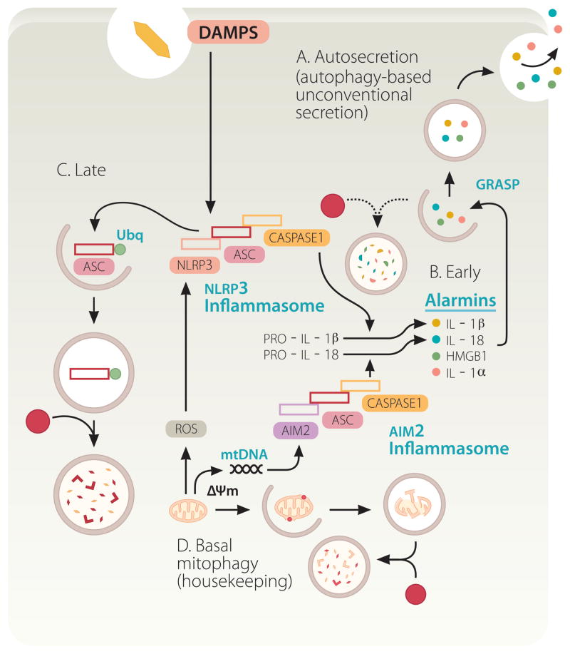
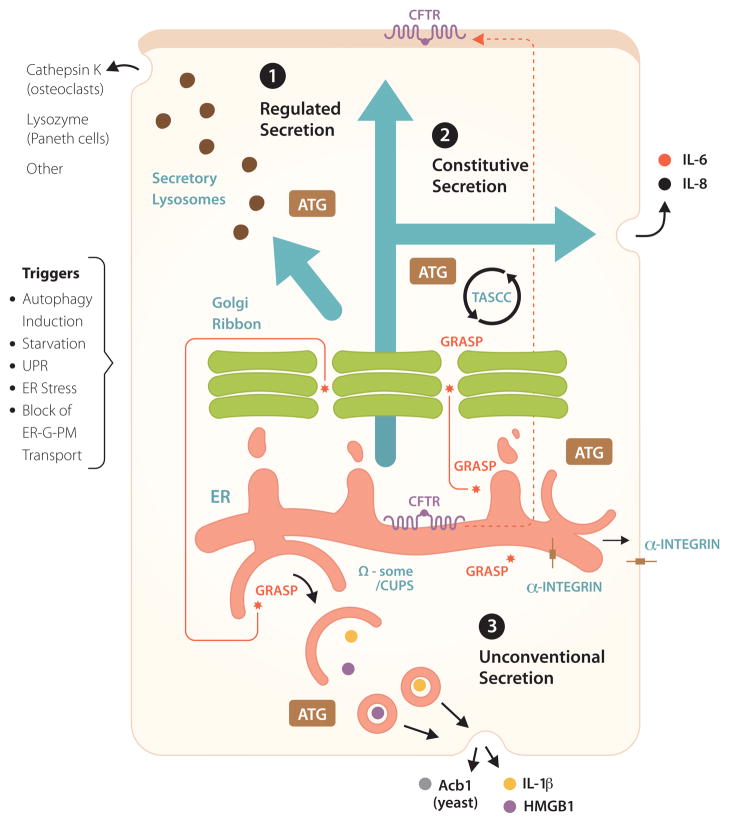
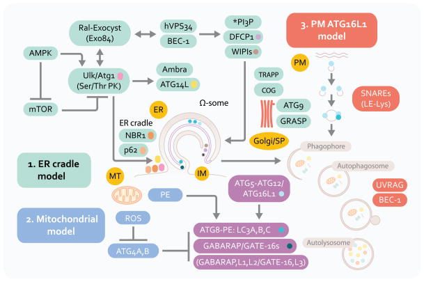
Autophagy is a cell biological process ubiquitous to all eukaryotic cells, often referred to as a catabolic, lysosomal degradative pathway. However, current studies in mammalian systems suggest that autophagy plays an unexpectedly broad biogenesis role in protein trafficking and secretion. Autophagy supports alternative trafficking pathways for delivery of integral membrane proteins to the plasma membrane and affects secretion, including the constitutive, regulated and unconventional secretion pathways. Autophagy-based unconventional secretion, termed here 'autosecretion', is one of the pathways enabling leaderless cytosolic proteins to exit the cell without entering the endoplasmic reticulum (ER)-to-Golgi secretory pathway. In this review, we discuss the emerging underlying mechanisms of how autophagy affects different facets of secretion. We also describe the physiological roles of autosecretory cargos that are often associated with inflammatory processes and also play a role in the formation of specialized tissues and in tissue remodeling, expanding the immediate sphere of influence of autophagy from the intracellular to the extracellular space.


Figures

Fig. 1
Fig. 1
Autophagy pathway: signaling systems and three proposed membrane sources in autophagosome formation. ER, endoplasmic reticulum. PM, plasma membrane. MT, mitochondria. Model 1: The central membranous structure, omegasome (Ω-some) is derived from the ER (ER cradle model), and is believed to be an early precursor of autophagic isolation membranes (IM) or phagophores. Phagophore crescents close to form double membrane autophagosomes that fuse with lysosomal intermediates to form the degradative organelles, autolysosomes. Model 2: Mitochondria may contribute membrane or phosphatidylinositol (PE) of relevance for LC3 (A, B and C; and other Atg8 paralogs, GABARAP and GATE-16) C-terminal lipidation into the LC3-II, autophagic membrane-associated form. Mitochondria may also be a source of reactive oxygen species that inactivate ATG4, an LC3 dilipidating enzyme. Mitochondria are also one of the major target substrates for autophagic elimination. Model 3: Plasma membrane Atg16L1-positive (initially LC3-negative) vesicles may contribute to autophagic membrane growth. Factors in the left upper corner represent upstream signaling systems (AMPK, mammalian Tor [mTor], Ral) controlling induction of autophagy in response to nutritional and cellular energetics signals. Beclin 1 (BEC-1) and class III phosphatidylinositol 3-kinase hVPS34 cooperate in control of phosphatidylinositol 3-phosphate (PI3P) structures that start with Ω-some, identifiable by the marker DFCP-1 for which a functional role in autophagy is yet to be established. NBR1 and p62 (also known as sequestosome 1) are autophagic adaptor proteins that capture cargo and interact with LC3; p62 is also present very early at the sites leading to omegasome formation, and is furthermore found in complexes with mTOR that sense amino acid starvation (not shown). Ambra and Atg14L are additional factors interacting with Beclin 1 complexes that are responsible for the early autophagosomal pathway. The lipid kinase hVPS34 interacts with UVRAG and additional factors (not shown) to control autophagosomal maturation into autolysosomes. Several systems and tethering systems along the different stages of the early secretory pathway and the Golgi apparatus (TRAPP, COG, GRASP) influence the formation, expansion (contributed by the only Atg integral membrane protein Atg9) and maturation of autophagosomal organelles.
Fig. 2
Fig. 2
Non-degradative roles of autophagy in conventional (constitutive and regulated) and unconventional secretion. 1. Regulated secretion: secretory lysosomes, granules and other organelles, partially derived from or affected by post-Golgi vesicles. ATG: symbolizes that Atg factors affect regulated secretion, delivering various biologically active cargo such as indicated. Other: includes non-proteinaceous cargo (e.g. ATP secreted from drug-treated cancer cells), provided that they are competent to undergo autophagy, with inflammatory consequences and clearance of transplanted tumors. 2. Autophagy affects constitutive secretion (e.g. IL-6, IL-8) via a compartment intermixed with autophagic organelles, called TASCC (TOR-autophagy spatial coupling compartment). 3. A subset of unconventional secretion processes depend on autophagy (autophagy-based unconventional secretion; ‘autosecretion’) for secretion of proinflammatory factors IL-1β and HMGB1 in mammalian cells and Acb1 in yeast. GRASP (note in Figure 1 that GRASP is normally localized to the Golgi and affects early stages of autophagy) is required for autophagy-based unconventional secretion (autosecretion). CUPS, a yeast structure implicated in autophagy-based unconventional secretion, may be equivalent to Ω-some in mammalian cells. In addition, autophagy plays a role in unconventional trafficking of the ER-form of CFTR (cystic fibrosis transmembrane conductance regulatory) to the apical aspect of the plasma membrane, bypassing the Golgi and rescuing function of mutant CFTR responsible for cystic fibrosis. GRASP plays a role in autophagy-dependent unconventional trafficking of CFTR and in unconventional trafficking of α-integrin to the basolateral plasma membrane in Drosophila (a role for autophagy has not been established as yet for α-integrin trafficking). Triggers: conditions (the list is not comprehensive) that contribute to induction of unconventional secretion or trafficking.
Fig. 3
Fig. 3
Autophagy plays a dual role in pro-inflammatory processes based on inflammasome activation and autosecretion. Autophagy suppresses basal levels of inflammasome activation. Inflammasomes (composed of an NLR, in the illustrated case NLRP3, or AIM2, ASC, and caspase 1) are multi-protein platforms assembled and activated in response to damage/danger associated molecular patterns (DAMPS), resulting in caspase-1 activation and proteolytic processing of cytosolic pro-forms of cytokines such as IL-1β, a cardinal mediator of infection-associated and sterile inflammation. Autophagy keeps the sources of endogenous agonists low by removing depolarized (ΔΨm) or unkempt mitochondria (sources of ROS and mitochondrial DNA [mtDNA]). However, autophagy also enables the cytosolic IL-1β (and other alarmins such as HMGB1; see Figure 2) to be delivered to the outside of the cell via autosecretion. Finally, autophagy may downregulate inflammasomes by digesting its components. A, B. Autosecretion (autophagy-based unconventional secretion; see Figure 2 and text for explanations) enables secretion of cytosolic proteins such as IL-1β and HMGB1 per the illustrated process controlled by Atg factors and GRASP. Autosecretion occurs early in the process of stimulation. C. Autosecretion is likely shut down by deactivation, or possibly by digestion as in the illustration, as a way of downregulating the system following its physiologically useful period of activation. D. In resting cells, the effect of autosecretion is masked by the dominant tonic negative role of autophagy in inhibiting basal levels of inflammasome activation.

References

    1. Mizushima N, et al. The role of atg proteins in autophagosome formation. Annual review of cell and developmental biology. 2011;27:107–132. - PubMed
    1. Liu L, et al. Mitochondrial outer-membrane protein FUNDC1 mediates hypoxia-induced mitophagy in mammalian cells. Nat Cell Biol. 2012;14:177–185. - PubMed
    1. Youle RJ, Narendra DP. Mechanisms of mitophagy. Nat Rev Mol Cell Biol. 2011;12:9–14. - PMC - PubMed
    1. Green DR, et al. Mitochondria and the autophagy-inflammation-cell death axis in organismal aging. Science. 2011;333:1109–1112. - PMC - PubMed
    1. Rubinsztein DC, et al. Autophagy and aging. Cell. 2011;146:682–695. - PubMed

Publication types