. 2012 Jun 15;26(12):1312-25.
doi: 10.1101/gad.191866.112. Epub 2012 Jun 7.

Scube/You activity mediates release of dually lipid-modified Hedgehog signal in soluble form

Affiliations

Scube/You activity mediates release of dually lipid-modified Hedgehog signal in soluble form

Adrian Creanga et al. Genes Dev. .

Abstract

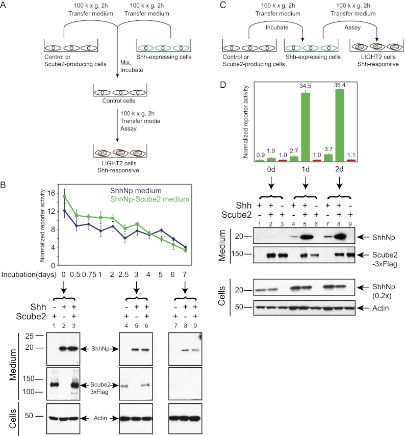
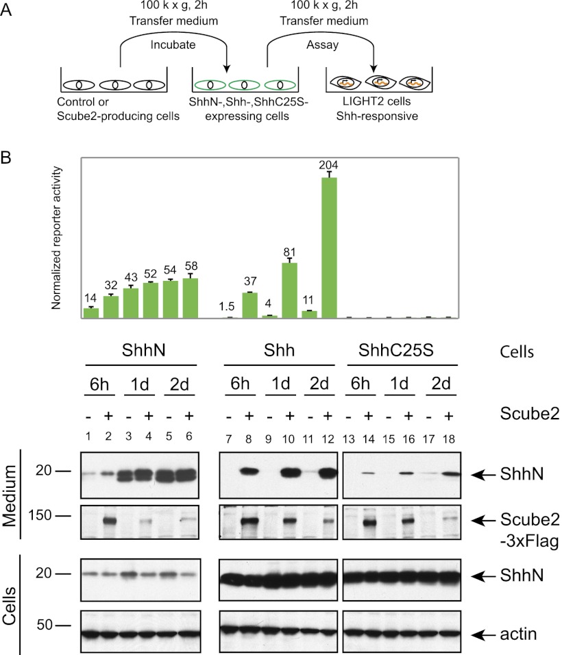
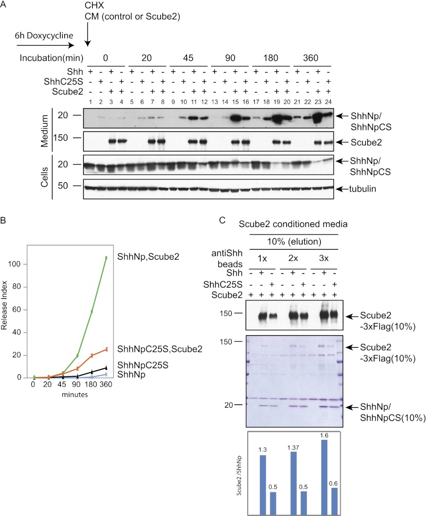
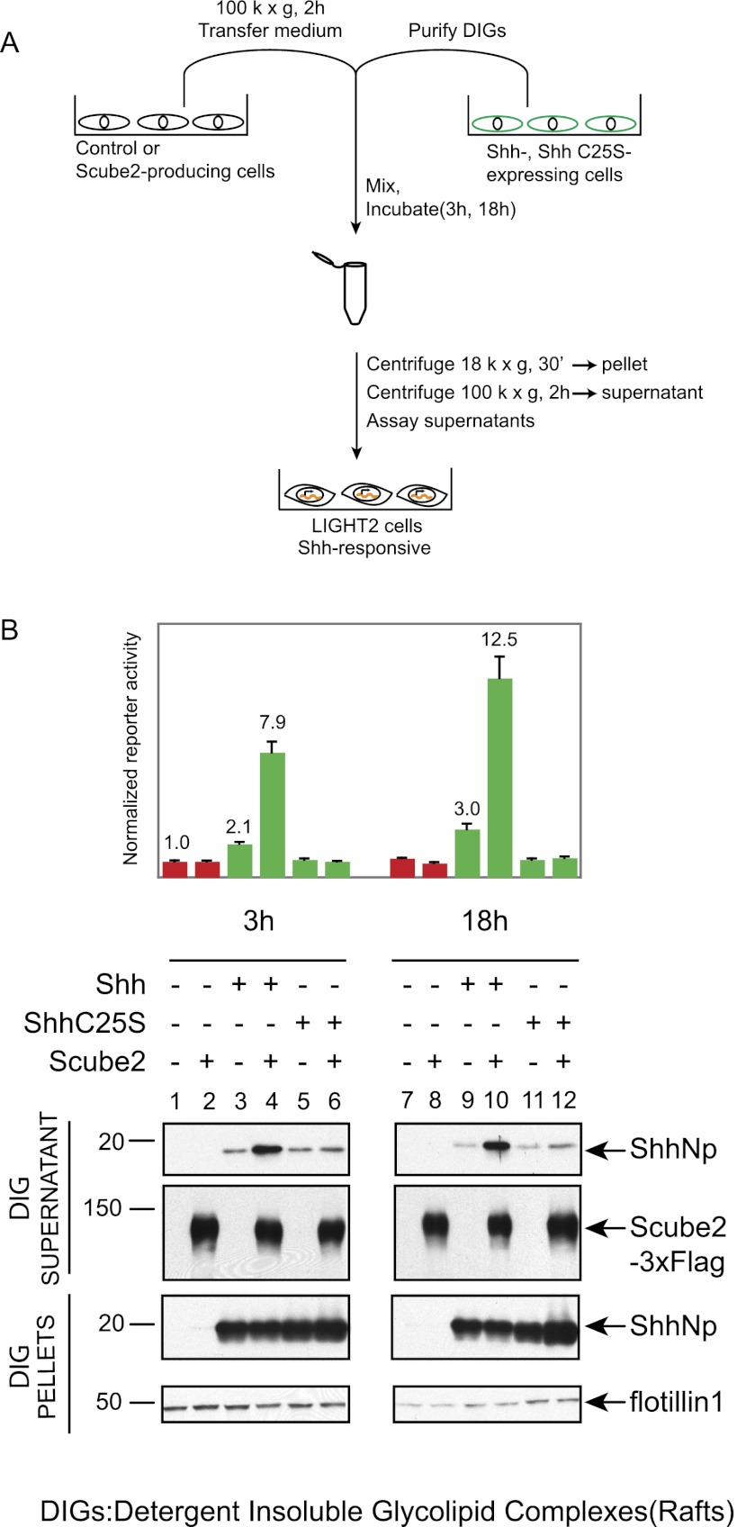
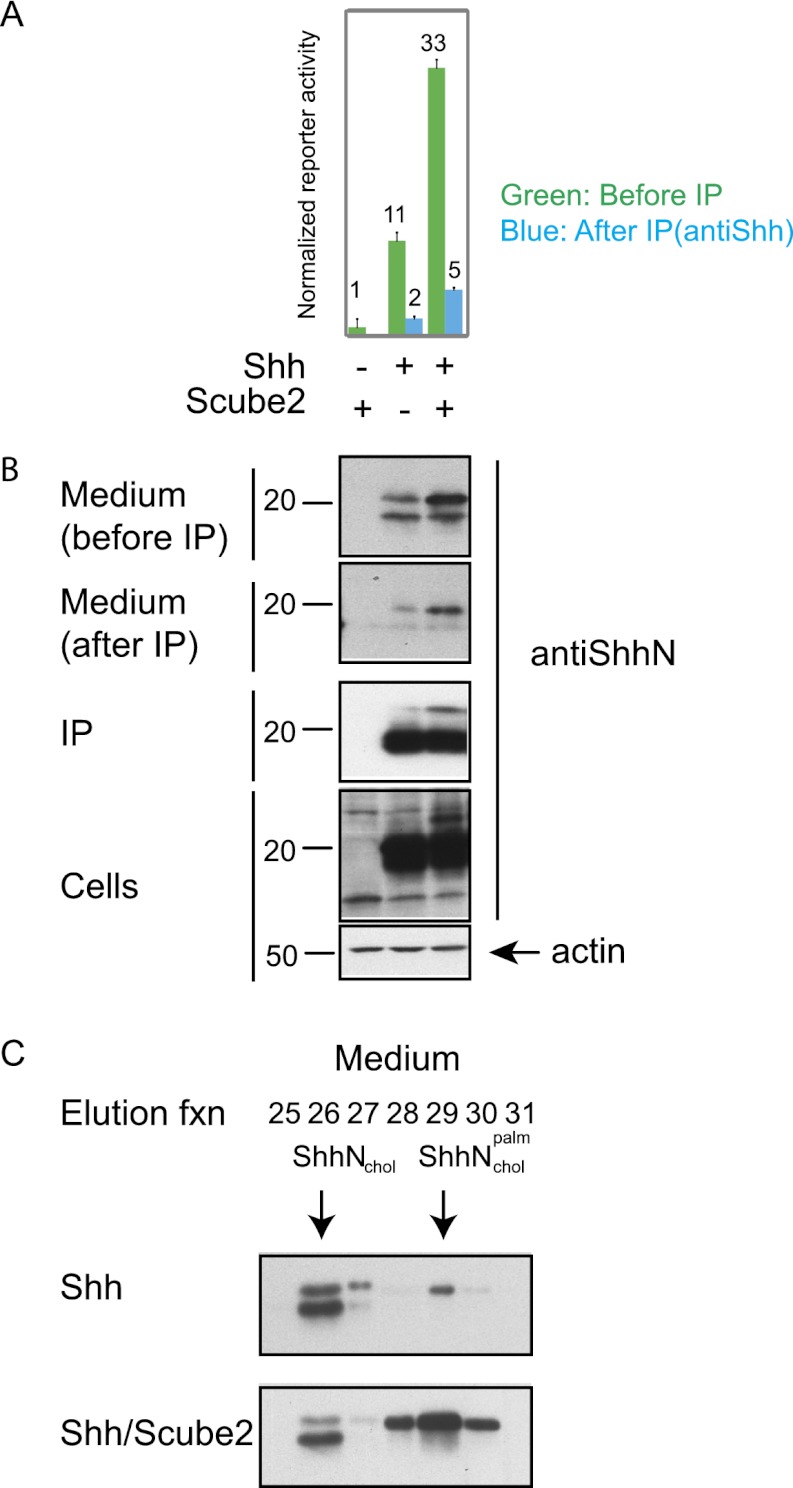
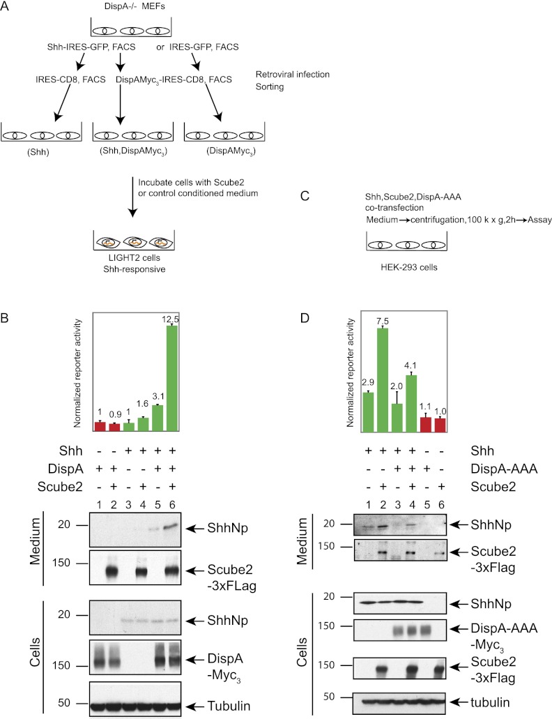
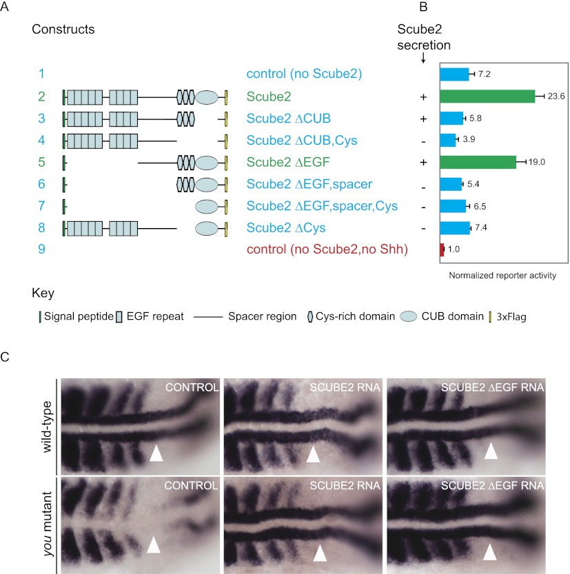
Owing to their covalent modification by cholesterol and palmitate, Hedgehog (Hh) signaling proteins are localized predominantly to the plasma membrane of expressing cells. Yet Hh proteins are also capable of mobilizing to and eliciting direct responses from distant cells. The zebrafish you gene, identified genetically >15 years ago, was more recently shown to encode a secreted glycoprotein that acts cell-nonautonomously in the Hh signaling pathway by an unknown mechanism. We investigated the function of the protein encoded by murine Scube2, an ortholog of you, and found that it mediates release in soluble form of the mature, cholesterol- and palmitate-modified Sonic hedgehog protein signal (ShhNp) when added to cultured cells or purified detergent-resistant membrane microdomains containing ShhNp. The efficiency of Scube2-mediated release of ShhNp is enhanced by the palmitate adduct of ShhNp and by coexpression in ShhNp-producing cells of mDispatchedA (mDispA), a transporter-like protein with a previously defined role in the release of lipid-modified Hh signals. The structural determinants of Scube2 required for its activity in cultured cell assays match those required for rescue of you mutant zebrafish embryos, and we thus conclude that the role of Scube/You proteins in Hh signaling in vivo is to facilitate the release and mobilization of Hh proteins for distant action.


Figures

Figure 1.
Figure 1.
Scube2 coexpression with Shh increases the release of soluble, active ShhNp protein. (A) Medium was harvested from HEK293S cells transiently transfected with expression plasmids for 3xFlag-tagged Scube2 or a control plasmid (see the Materials and Methods) along with Shh (2:1 ratio of Scube2 to Shh), and centrifuged at 18,000g for 30 min, and signaling was activity measured using Shh-responsive NIH-3T3 cells stably transfected with firefly luciferase-based Shh pathway reporter and Renilla luciferase constitutive reporter (Shh-LIGHT2 cells). Medium harvested 48 h post-transfection was centrifuged at 100,000g for an additional 2 h. (B) Signaling activities of ShhNp-conditioned media collected from transfected HEK293S cells at specified time points and incubated for 48 h with Shh-LIGHT2 cells at a fivefold dilution of conditioned media with DMEM containing 0.5% calf serum. Relative reporter activities (firefly luciferase/Renilla luciferase) were normalized to relative reporter activity produced by medium collected from HEK293 cells not transfected with the Shh-expressing construct (shown in red here and throughout the study). Culture media and whole-cell lysates harvested from HEK293S cells transiently transfected with the indicated constructs were probed by immunoblotting for expression of ShhNp (anti-ShhN antibody) and 3xFlag-tagged Scube2 (anti-Flag antibody). Tubulin was used as a loading control. Migration of molecular markers is indicated. (C) Scube2 increases the activity of ShhNp-conditioned medium by increasing the amount of soluble ShhNp in the medium. Medium collected 2 d after transfection and centrifuged at 18,000g for 30 min was further centrifuged at 100,000g for 2 h. The signaling activity of the medium was measured by incubation with Shh-LIGHT2 cells, and protein levels were analyzed by reducing SDS-PAGE and immunoblotting to detect Scube2-3xFlag (anti-Flag) and ShhNp proteins (anti-ShhN).
Figure 2.
Figure 2.
Externally supplied Scube2 acts by enhancing the release of ShhNp. (A) Experimental scheme to test for Scube2-mediated stabilization of ShhNp. HEK293 Flp-In T-REX cells with stably integrated constructs for tetracycline-inducible expression of Shh or 3xFlag-tagged Scube2 were incubated with doxycycline-containing medium. Medium harvested from these or control parental cells was centrifuged at 18,000g for 30 min followed by 100,000g for 2 h and mixed at a 1:1 ratio (v/v) to prepare three distinct conditioned media: (1) Scube2-conditioned medium, with 50% Scube2-conditioned medium and 50% control conditioned medium from parental cells; (2) ShhNp-conditioned medium, with 50% ShhNp-conditioned medium and 50% control conditioned medium; and (3) ShhNp–Scube2-conditioned medium, with 50% ShhNp-conditioned medium and 50% Scube2-conditioned medium. Each of these conditioned media was further incubated with parental HEK293 Flp-In cells for the indicated periods of time prior to assay. (B) Scube2 does not affect signaling activity or the stability of secreted ShhNp. Signaling activity of conditioned medium was measured at fivefold dilution with Shh-LIGHT2 cells. Signaling activity of control conditioned medium incubated with cells not expressing Shh was used for normalization. Media samples were also probed for ShhNp (anti-ShhN) and 3xFlag-tagged Scube2 (anti-Flag) by immunoblotting, with lysates from proportional fractions of cell cultures probed for actin expression as controls. (C) Experimental scheme to test for Scube2-mediated release of ShhNp. Stably integrated, tetracycline-inducible cells were used to prepare control or Scube2-containing medium as described in A. After 12 h of doxycycline treatment, medium from parental or Shh-expressing cells was replaced with control or Scube2-conditioned medium and incubated for the indicated periods of time. ShhNp-conditioned medium was centrifuged at 18,000g for 30 min, and the resulting supernatant was further centrifuged at 100,000g for 2 h. Activity measurements were carried out with Shh-LIGHT2 cells and 20-fold dilution of conditioned media with DMEM containing 0.5% calf serum. (D) Scube2 stimulates the release of ShhNp protein and signaling activity. Shh-expressing cells were incubated with control or Scube2-containing conditioned medium. Signaling activity of control conditioned medium was used for normalization. Scube2-containing conditioned medium increased signaling activity by ∼10-fold and also increased the release of ShhNp, as detected by immunoblotting (performed as described in B).
Figure 3.
Figure 3.
Role of lipid modification in Scube2-mediated release of the Shh signal. (A) HEK293 Flp-In T-REX cells were used to establish isogenic cell lines expressing ShhN, Shh, or ShhC25S. After 24 h of incubation with doxycycline-containing medium to induce protein expression, cells were incubated with control or Scube2-conditioned medium prepared as in Figure 2C and harvested after 6 h or 1 or 2 d of incubation. (B) Scube2 dramatically increases release of signaling activity from cells expressing ShhNp but not ShhN. The signaling activity of the medium was measured at 20-fold dilution using Shh-LIGHT2 cells. The signaling activity of the control conditioned medium was used for normalization. Immunoblotting of medium and whole-cell lysates using anti-ShhN (ShhN, ShhNp, or ShhNpC25S) and anti-Flag (Scube2-3xFlag) indicates that ShhN does not require Scube2 for its release in the medium and that palmitate modification augments Scube2-mediated release of ShhNp.
Figure 4.
Figure 4.
Palmitate modification of ShhNp enhances Scube2-mediated release and contributes to the interaction between ShhNp and Scube2. (A) Time course of Scube2-mediated ShhNp release from cells expressing Shh or ShhC25S. HEK293 Flp-In T-REX cells with stably integrated constructs for tetracycline-inducible expression of Shh or ShhC25S were treated for 6 h with doxycycline, and medium was replaced with control or Scube2-conditioned medium containing 50 μg/mL cycloheximide. Medium and whole-cell lysates harvested at the indicated time points were immunoblotted with anti-ShhN antibody (ShhN) and anti-Flag (Scube2-3xFlag), with tubulin as a loading control. The migration of the molecular markers is indicated. (B) Autoradiograms in A were quantified using ImageJ, and the release index ([ShhNp]medium/[ShhNp]cells) was plotted for ShhNp and ShhNpC25S in the presence or absence of externally added Scube2. (C) The palmitoyl adduct of ShhNp increases binding of Scube2. Control, Shh-expressing, or Shh C25-expressing HEK293 Flp-In T-Rex cells were lysed in radioimmunoprecipitation assay (RIPA) buffer (see the Materials and Methods), and lysates were incubated for 12 h with increasing quantities (1×, 2×, and 3×) of anti-Shh antibody (5E1) immobilized on Affigel 10 beads. Beads were washed and incubated for 4 h with Scube2-containing medium, and precipitated proteins were eluted with denaturing sample buffer and analyzed by SDS/PAGE followed by immunoblotting (top panel) or Coomassie staining (middle panel). (Bottom panel) Quantification of Scube2-3xFlag and ShhNp/ShhNpC25S from scanned, Coomassie-stained gels with ImageJ software showed that Scube2 is more efficiently precipitated from medium by beads loaded with ShhNp as compared with ShhNpC25S.
Figure 5.
Figure 5.
Scube2 increases the release of ShhNp from lipid rafts in a cell-free assay. (A) Experimental scheme to test for Scube2-mediated release of ShhNp protein in a cell-free assay. Lipid rafts (DIGs) isolated from HEK293 Flp-In T-REX cells expressing Shh or ShhC25S were purified by sucrose gradient fractionation and incubated with control or Scube2-containing conditioned medium prepared without serum for the indicated periods of time. (B) Signaling activities of supernatants obtained by incubation of ShhNp- or ShhNpC25S-containing lipid rafts with control or Scube2-containing conditioned medium for 3 h or 18 h and centrifuged at 100,000g for 2 h were analyzed with Shh-LIGHT2 cells at threefold dilution. DIG pellets and supernatants were probed for ShhNp (anti-ShhN) and Scube2-3xFlag (anti-Flag) by immunoblotting, with DIG pellets probed also for Flotillin-1 as loading controls. The migration of the molecular markers is indicated. Scube2-containing conditioned medium increases the signaling activity of ShhNp supernatants up to fourfold, and the presence of palmitate modification increases the Scube2-mediated release of ShhNp protein from DIGs in a cell-free assay.
Figure 6.
Figure 6.
Soluble ShhNp released by Scube2 is dually lipid-modified. (A) HEK293S cells transiently transfected (24 h) with expression plasmids for 3xFlag-tagged Scube2 or control plasmid along with Shh (2:1 ratio) were incubated for 4 d with medium lacking transfection reagent. After harvesting, medium was centrifuged at 100,000g for 2 h and incubated with immobilized anti-ShhN (5E1 antibody linked to Affigel 10 beads) for 24 h. The signaling activity of the medium before (green) and after (blue) incubation with anti-ShhN beads was analyzed by incubation with Shh-LIGHT2 cells at fivefold dilution. Note that the signaling activity is depleted by incubation with anti-ShhN beads. (B) Culture medium before and after incubation with anti-ShhN beads, immunoprecipitated protein, and whole-cell lysate (with actin as a loading control) was immunoblotted for ShhNp (anti-ShhN). Note the efficiency of ShhNp depletion from the medium. (C) Lipid modification of ShhNp as determined by reverse-phase HPLC. ShhNp immunoprecipitated from conditioned medium as in B was analyzed by reverse-phase HPLC in a C4 column developed with a gradient of increasing acetonitrile in water containing 0.1% trifluoroacetic acid to determine the degree of ShhN lipid modification. Fractions eluted from reverse-phase HPLC were analyzed by immunoblotting after vacuum-drying and resolubilization with 1× protein sample buffer. Representative elution fractions are shown, and elution standards were characterized by reverse-phase HPLC analysis of ShhNp, ShhNpC25S, ShhN, and ShhNC25S proteins (Supplemental Fig. 3). ShhNp-conditioned medium prepared in the presence of Scube2 contains increased levels of dually lipidated ShhNp (formula image)
Figure 7.
Figure 7.
Scube2-mediated release of ShhNp is facilitated by Dispatched. (A) Experimental scheme to test for the role of DispA in the Scube2-mediated release of ShhNp. DispA−/− MEF cells transduced with a MIGR1 retrovirus-based construct expressing Shh-IRES-GFP or IRES-GFP were FACS-sorted for GFP. Next, GFP+ cells were transduced with DispA-IRES-CD8 or IRES-CD8 and FACS-sorted for CD8. These cells were incubated with control or Scube2-containing conditioned medium for 2 d. (B) Scube2 stimulates the release of ShhNp protein and signaling activity only in the presence of DispA. The signaling activity of media collected from DispA−/− cells transduced with the indicated expression constructs and incubated with Scube2- or control conditioned medium was analyzed with Shh-LIGHT2 cells at twofold dilution. Media and whole-cell extracts were probed for ShhNp (anti-ShhN), 3xFlag-tagged Scube2 (anti-Flag), and 3xMyc-tagged DispA (anti-Myc) by immunoblotting, with tubulin used as a loading control. The migration of the molecular markers is indicated. In the presence of DispA, Scube2-containing conditioned medium increased signaling activity by approximately fourfold and increased the level of released ShhNp detectable by immunoblotting. (C) Experimental scheme to test effect of the DispA-AAA dominant-negative. HEK293S cells cultured in six-well plates were transiently transfected with 1:6:15 (w/w/w) ratios of plasmids expressing Shh, Scube2, and DispA-AAA, respectively. The signaling activities of medium collected 4 d post-transfection were analyzed by incubation with Shh-LIGHT2 cells. (D) Scube2-mediated release of ShhNp in the medium is reduced by expression of DispA-AAA. Signaling activities of media samples were measured with Shh-LIGHT2 cells at fivefold dilution. Culture medium and whole-cell lysates were probed by immunoblotting for expression of ShhNp (anti-ShhN antibody), 3xFlag-tagged Scube2 (anti-Flag antibody), and 3xMyc-tagged DispA-AAA (anti-Myc antibody). Note that the signaling activity and the release of ShhNp into the medium were decreased by coexpression of DispA-AAA.
Figure 8.
Figure 8.
Structure–function analysis of Scube2 function in vitro and in vivo. (A) Schematic diagram shows 3xFlag-tagged Scube2 deletion constructs used for this analysis. (B) Signaling activity of medium from HEK293S cells transiently cotransfected with expression plasmids for Shh and wild-type or deleted 3×Flag-tagged Scube2 (1:2 ratio) was measured with Shh-LIGHT2 cells at fivefold dilution. For immunoblotting analysis of these samples, see Supplemental Figure 4. Secretion of Scube2 protein variants into culture medium as detected by immunoblotting is indicated (+ or −). (C) Rescue of you mutants by microinjection of zscube2 mRNA constructs. (Top row) Wild-type embryos show adaxial expression of myod (white arrowheads). (Bottom left) You mutant embryos lack adaxial myod expression (white arrowhead). Injection of full-length zscube2 mRNA (middle column) and zscube2ΔEGF mRNA (right column) rescues the expression of myod in adaxial cells in you mutants (white arrowheads).

Comment in

References

    1. Beachy PA, Karhadkar SS, Berman DM 2004. Tissue repair and stem cell renewal in carcinogenesis. Nature 432: 324–331 - PubMed
    1. Briscoe J, Chen Y, Jessell TM, Struhl G 2001. A hedgehog-insensitive form of patched provides evidence for direct long-range morphogen activity of sonic hedgehog in the neural tube. Mol Cell 7: 1279–1291 - PubMed
    1. Buglino JA, Resh MD 2008. Hhat is a palmitoylacyltransferase with specificity for N-palmitoylation of Sonic Hedgehog. J Biol Chem 283: 22076–22088 - PMC - PubMed
    1. Burke R, Nellen D, Bellotto M, Hafen E, Senti KA, Dickson BJ, Basler K 1999. Dispatched, a novel sterol-sensing domain protein dedicated to the release of cholesterol-modified hedgehog from signaling cells. Cell 99: 803–815 - PubMed
    1. Caspary T, Garcia-Garcia MJ, Huangfu D, Eggenschwiler JT, Wyler MR, Rakeman AS, Alcorn HL, Anderson KV 2002. Mouse Dispatched homolog1 is required for long-range, but not juxtacrine, Hh signaling. Curr Biol 12: 1628–1632 - PubMed

MeSH terms

LinkOut - more resources