Clinical Trial
. 2012 Jul;23(7):1229-37.
doi: 10.1681/ASN.2011121186. Epub 2012 Jun 7.

Pathology after eculizumab in dense deposit disease and C3 GN

Affiliations
Clinical Trial

Pathology after eculizumab in dense deposit disease and C3 GN

Leal C Herlitz et al. J Am Soc Nephrol. 2012 Jul.

Abstract

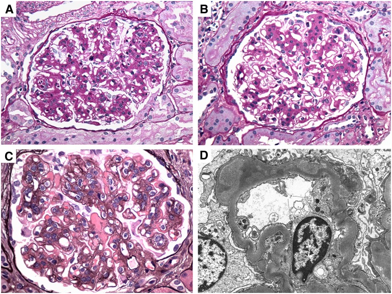
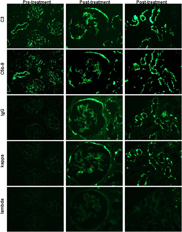
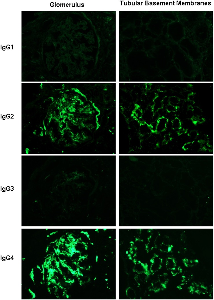
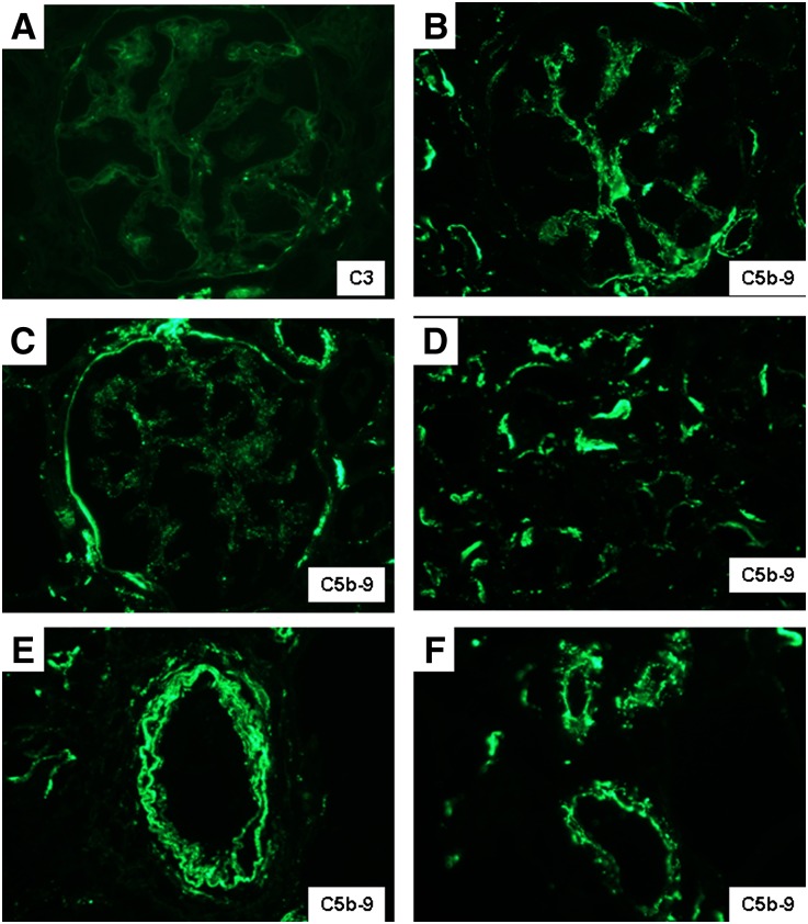
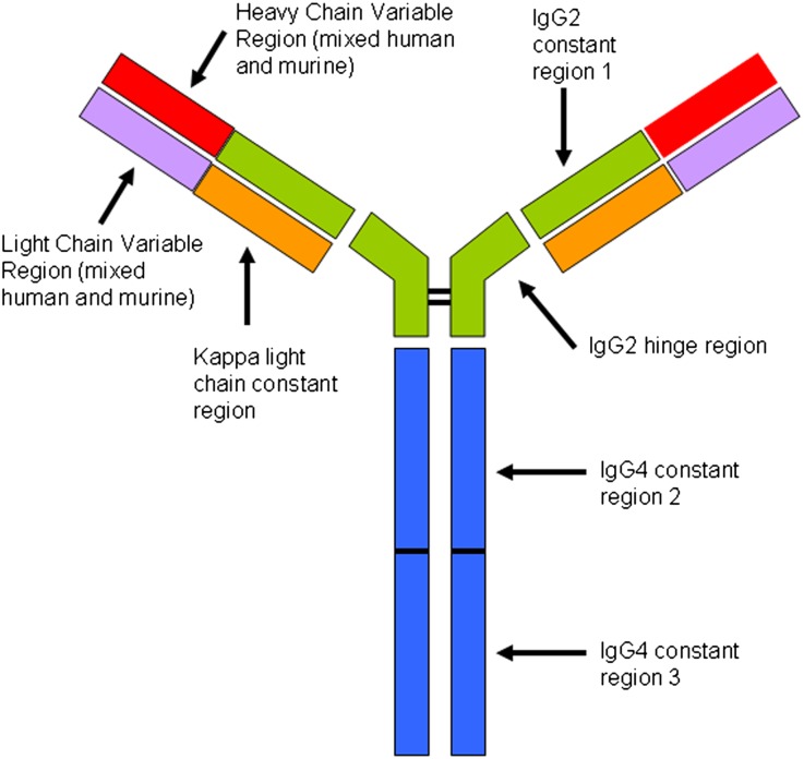
Eculizumab might benefit C3 glomerulopathies mediated by dysregulation of the alternative complement pathway. Here, we report renal biopsy findings before and after eculizumab therapy in three patients with dense deposit disease and two with C3 GN. All pretreatment biopsies had glomerular and tubular basement membrane deposits that stained exclusively for C3 without significant Ig. After 1 year of therapy, there was reduction in active glomerular proliferation and neutrophil infiltration in three of five patients, consistent with effective C5 blockade, which prevents production of chemotactin C5a. One individual with mild mesangial disease had no significant change in activity or chronicity. One patient exhibited persistent activity and worsening chronicity despite therapy. Immunofluorescence showed no significant reduction in C3 or C5b-9, and electron microscopy revealed persistent deposits in all cases, suggesting a long t(1/2) of C5b-9 in extracellular matrix. Normal renal biopsies stained positive for C5b-9 in glomeruli, tubular basement membranes, and vessel walls, albeit at lower intensity than in C3 glomerulopathy. This indication of physiologic levels of C5b-9 activation in normal kidney potentially explains the localization of deposits in patients with dysregulation of the alternative complement pathway. All post-treatment biopsies showed de novo monoclonal staining for IgG-κ in the same distribution as C3 and C5b-9, mimicking monoclonal Ig deposition disease (MIDD). Staining of the γ heavy chain was restricted to the IgG2 and IgG4 subclasses, suggesting the binding of monoclonal eculizumab to C5 in renal tissues. The long-term effects of this apparent drug-tissue interaction are unknown.


Figures

Figure 1.
Figure 1.
Representative pretreatment and post-treatment histologic and ultrastructural findings in C3 glomerulpathy. (A) Renal biopsies from patient DDD1 show segmental endocapillary proliferation and leukocyte infiltration on a base of mesangial proliferation and sclerosis in the pretreatment biopsy (periodic acid–Schiff). (B) After treatment, there is resolution of the active features of endocapillary proliferation and leukocyte exudation, but with persistent mesangial proliferation (periodic acid–Schiff). (C) Patient C3GN1, by contrast, has persistent membranoproliferative features with large subendothelial deposits post-treatment (Jones methenamine silver). (D) Patient DDD1 exhibits a mild decrease in the electron density of the intramembranous deposits post-treatment (electron micrograph). Original magnification, ×600 in A through C; ×10,000 in D.
Figure 2.
Figure 2.
A representative panel of serial sections from patient C3GN3 illustrates the evolution of staining for C3, C5b-9, IgG, κ light chain, and λ light chain in pretreatment and post-treatment biopsies. Column 1 shows pretreatment findings in a low-power (×200) view of three glomeruli and an interlobular artery, which demonstrate positivity for C3 and C5b-9, but no staining for IgG or either light chain. Columns 2 (×400; glomerulus) and 3 (×600; tubules) illustrate post-treatment biopsy findings, with the persistence of C3 and C5b-9 in glomerular tufts, Bowman’s capsules, and TBMs, as well as the new appearance of positivity for IgG and κ (with negative λ) in the same distribution.
Figure 3.
Figure 3.
Immunofluorescence staining in serial sections for the IgG subtypes in the post-treatment biopsy of patient DDD1 shown for glomeruli (×600; column 1) and tubules (×400; column 2) reveals positivity for IgG2 and IgG4, with negativity for IgG1 and IgG3 in the distribution of the IgG-κ deposits. Similar results were seen in all of the post-treatment biopsies examined.
Figure 4.
Figure 4.
Representative images of the post-treatment biopsy from patient C3GN1 illustrate strong glomerular staining for IgG and κ light chain, with negativity for λ light chain (column 1; ×600). By electron microscopy, the electron dense subendothelial deposits exhibited an unusual punctate highly electron dense powdery texture, resembling those seen in MIDD. This transformation in the appearance of the deposits post-therapy may represent binding of eculizumab to existing deposits. Original magnification, ×15,000 in A; 40,000 in B.
Figure 5.
Figure 5.
Representative distribution of C5b-9 in normal human renal biopsies is illustrated. (A) In contrast to the weak and sparse mesangial staining for C3, (B) the identical field stained for C5b-9 reveals stronger mesangial positivity, in addition to staining of the preglomerular arteriolar wall. Focal staining of (C) Bowman’s capsule and (D) TBMs and diffuse staining of (E) arterial walls and (F) arteriolar walls are also observed. Original magnification, ×600 in A through C and F; 400 in D and E.
Figure 6.
Figure 6.
Structural features of eculizumab. Adapted from Rother et.al. Constant regions CH2 and CH3 from IgG4 are fused to the IgG2 hinge region and CH1 domain and then paired with a κ light chain. The variable regions of the light and heavy chains are composed of the murine-derived sequences that have high affinity for C5, admixed with human germline framework regions. The result is a hybrid Ig with high affinity for C5 but without the ability to activate complement or bind Fc receptors. Post-treatment biopsies show evidence of eculizumab binding to glomeruli, TBMs, and vessel walls in the form of immunofluorescence staining for IgG2, IgG4, and κ light chain, with negative staining for IgG1, IgG3, and λ light chain.

References

    1. Fakhouri F, Frémeaux-Bacchi V, Noël LH, Cook HT, Pickering MC: C3 glomerulopathy: A new classification. Nat Rev Nephrol 6: 494–499, 2010 - PubMed
    1. Nasr SH, Valeri AM, Appel GB, Sherwinter J, Stokes MB, Said SM, Markowitz GS, D’Agati VD: Dense deposit disease: Clinicopathologic study of 32 pediatric and adult patients. Clin J Am Soc Nephrol 4: 22–32, 2009 - PMC - PubMed
    1. Servais A, Frémeaux-Bacchi V, Lequintrec M, Salomon R, Blouin J, Knebelmann B, Grünfeld JP, Lesavre P, Noël LH, Fakhouri F: Primary glomerulonephritis with isolated C3 deposits: A new entity which shares common genetic risk factors with haemolytic uraemic syndrome. J Med Genet 44: 193–199, 2007 - PMC - PubMed
    1. D'Agati VD, Jennette JC, Silva FG: Non-Neoplastic Kidney Diseases, Washington, DC, American Registry of Pathology, 2005, pp 250–255
    1. Sethi S, Gamez JD, Vrana JA, Theis JD, Bergen HR, 3rd, Zipfel PF, Dogan A, Smith RJ: Glomeruli of dense deposit disease contain components of the alternative and terminal complement pathway. Kidney Int 75: 952–960, 2009 - PMC - PubMed

Publication types

MeSH terms

LinkOut - more resources