

Effect of glucocorticoid (triamcinolone acetonide) pretreatment in a murine penetrating keratoplasty and suture model
- PMID: 22677639
- PMCID: PMC3443542
- DOI: 10.1097/ICO.0b013e3182473356
Effect of glucocorticoid (triamcinolone acetonide) pretreatment in a murine penetrating keratoplasty and suture model
Abstract
Purpose: To evaluate the effect of glucocorticoid (triamcinolone acetonide injectable suspension) pretreatment on corneal neovascularization, lymphangiogenesis, and inflammation in a murine penetrating keratoplasty (PK) and corneal suture model.
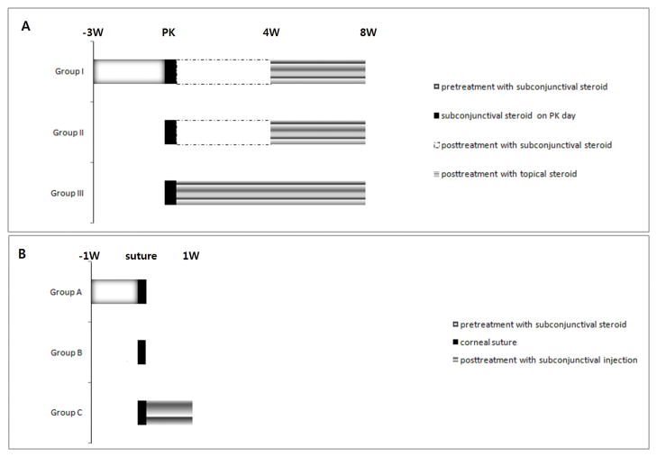
Methods: For the PK model, BALB/c mice were used as recipients and C57BL/6 mice were used as donors. A group pretreated with subconjunctival glucocorticoid and a combination of post-subconjunctival and topical glucocorticoids (group I) was compared with two groups that did not receive glucocorticoid pretreatment [one group received a combination of subconjunctival and topical glucocorticoids postoperatively (group II) and the other group received only topical glucocorticoid treatment postoperatively (group III)]. All groups were treated with subconjunctival glucocorticoid on the day of surgery. For the corneal suture model, BALB/c mice were used. A group receiving only pre-suture glucocorticoid treatment (group A) and a group receiving only post-suture glucocorticoid treatment (group C) were compared with a control group that did not receive glucocorticoid therapy (group B). The degree of neovascularization, lymphangiogenesis, and inflammatory infiltration was compared in each of these models.
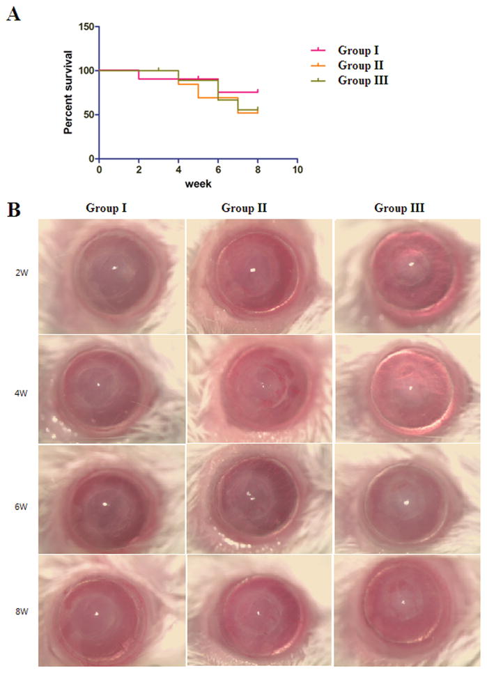
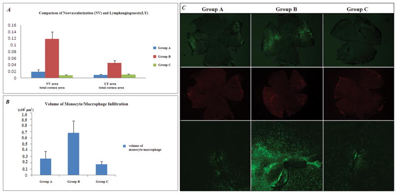
Results: In the PK model, the group receiving glucocorticoid pretreatment (group I) showed less neovascularization compared with the posttreatment-only groups (group II, P=0.043; group III, P=0.020) and less lymphangiogenesis compared with group III (P=0.005). In the corneal suture model, the glucocorticoid pretreatment group showed a similar level of neovascularization, lymphangiogenesis, and inflammatory infiltration as the posttreatment-only groups (P>0.05).
Conclusions: Glucocorticoid pretreatment before PK decreases neovascularization and lymphangiogenesis compared with posttransplant glucocorticoid treatment alone.
Conflict of interest statement
Dr. Ambati has received grant funding from the National Institutes of Health (NIH), is an employee of the University of Utah, and is on the speakers bureau for Alcon. All the authors are employees of University of Utah.
Figures

Similar articles
-
Vascular Endothelial Growth Factor Receptor 1 morpholino increases graft survival in a murine penetrating keratoplasty model.Invest Ophthalmol Vis Sci. 2012 Dec 19;53(13):8458-71. doi: 10.1167/iovs.12-10408. Invest Ophthalmol Vis Sci. 2012. PMID: 23150613 Free PMC article.
-
Flt23k nanoparticles offer additive benefit in graft survival and anti-angiogenic effects when combined with triamcinolone.Invest Ophthalmol Vis Sci. 2012 Apr 30;53(4):2328-36. doi: 10.1167/iovs.11-8393. Invest Ophthalmol Vis Sci. 2012. PMID: 22427553 Free PMC article.
-
Inflammatory corneal (lymph)angiogenesis is blocked by VEGFR-tyrosine kinase inhibitor ZK 261991, resulting in improved graft survival after corneal transplantation.Invest Ophthalmol Vis Sci. 2008 May;49(5):1836-42. doi: 10.1167/iovs.07-1314. Invest Ophthalmol Vis Sci. 2008. PMID: 18436817
-
[Anti(lymph)angiogenic preconditioning prior to keratoplasty].Klin Monbl Augenheilkd. 2013 May;230(5):500-4. doi: 10.1055/s-0032-1328500. Epub 2013 May 21. Klin Monbl Augenheilkd. 2013. PMID: 23695846 Review. German.
-
Subconjunctival triamcinolone acetonide in the management of ocular inflammatory disease.J Ocul Pharmacol Ther. 2013 Jul-Aug;29(6):516-22. doi: 10.1089/jop.2012.0208. Epub 2013 Mar 13. J Ocul Pharmacol Ther. 2013. PMID: 23485045 Review.
Cited by
-
Soluble vascular endothelial growth factor receptor 3 is essential for corneal alymphaticity.Blood. 2013 May 16;121(20):4242-9. doi: 10.1182/blood-2012-08-453043. Epub 2013 Mar 8. Blood. 2013. PMID: 23476047 Free PMC article.
-
Lymphatic vessels identified in failed corneal transplants with neovascularisation.Br J Ophthalmol. 2019 Mar;103(3):421-427. doi: 10.1136/bjophthalmol-2018-312630. Epub 2018 Oct 22. Br J Ophthalmol. 2019. PMID: 30348644 Free PMC article.
-
Effect of trapping vascular endothelial growth factor-A in a murine model of dry eye with inflammatory neovascularization.Int J Ophthalmol. 2016 Nov 18;9(11):1541-1548. doi: 10.18240/ijo.2016.11.02. eCollection 2016. Int J Ophthalmol. 2016. PMID: 27990354 Free PMC article.
-
Presurgical corticosteroid treatment improves corneal transplant survival in mice.Cornea. 2013 Dec;32(12):1591-1598. doi: 10.1097/ICO.0b013e31829ebb0d. Cornea. 2013. PMID: 24005616 Free PMC article.
References
-
- Lam H, Dana MR. Corneal graft rejection. Int Ophthalmol Clin. 2009 Winter;49(1):31–41. - PubMed
-
- The collaborative corneal transplantation studies (CCTS) Effectiveness of histocompatibility matching in high-risk corneal transplantation. The Collaborative Corneal Transplantation Studies Research Group. Arch Ophthalmol. 1992 Oct;110(10):1392–1403. - PubMed
-
- Streilein JW, Yamada J, Dana MR, Ksander BR. Anterior chamber-associated immune deviation, ocular immune privilege, and orthotopic corneal allografts. Transplant Proc. 1999 May;31(3):1472–1475. - PubMed
-
- Santos LN, de Moura LR, Fernandes BF, Cheema DP, Burnier MN., Jr Histopathological study of delayed regraft after corneal graft failure. Cornea. 2011 Feb;30(2):167–170. - PubMed
-
- Baradaran-Rafii A, Karimian F, Javadi M, et al. Corneal Graft Rejection: Incidence and Risk Factors. Iran J Ophthalmic Res. 2007;2(1):7–14.
Publication types
MeSH terms
Substances
Grants and funding
LinkOut - more resources
Full Text Sources
Medical
