. 2012 Jul 10;107(2):352-9.
doi: 10.1038/bjc.2012.251. Epub 2012 Jun 7.

miR-21, miR-17 and miR-19a induced by phosphatase of regenerating liver-3 promote the proliferation and metastasis of colon cancer

Affiliations

miR-21, miR-17 and miR-19a induced by phosphatase of regenerating liver-3 promote the proliferation and metastasis of colon cancer

J Zhang et al. Br J Cancer. .

Retraction in

Abstract

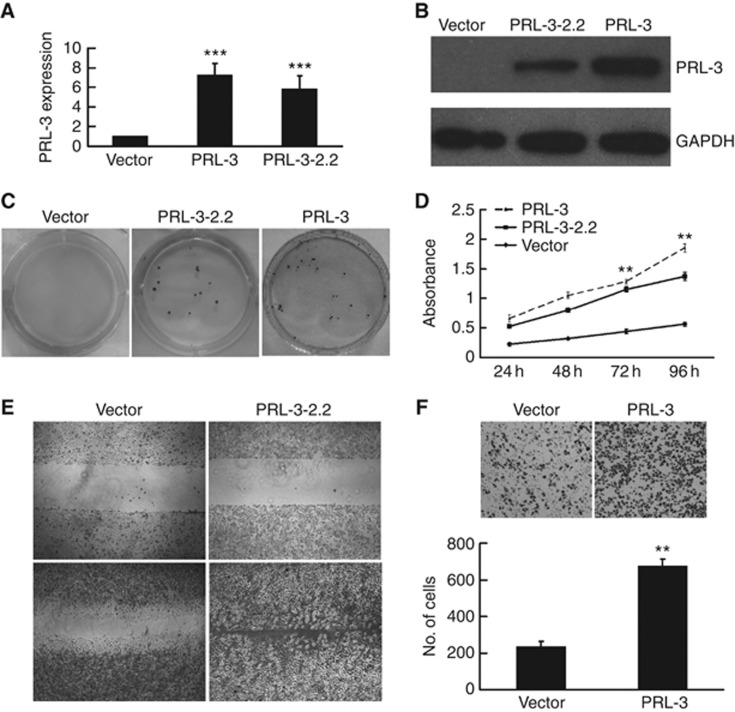
Background: Phosphatase of regenerating liver-3 (PRL-3) is an oncogene known to promote tumour metastasis, especially in colorectal cancer (CRC). Here, we demonstrate that the miR-21, miR-17 and miR-19a expressions induced by PRL-3 are involved in the proliferation and metastasis of colon cancer.

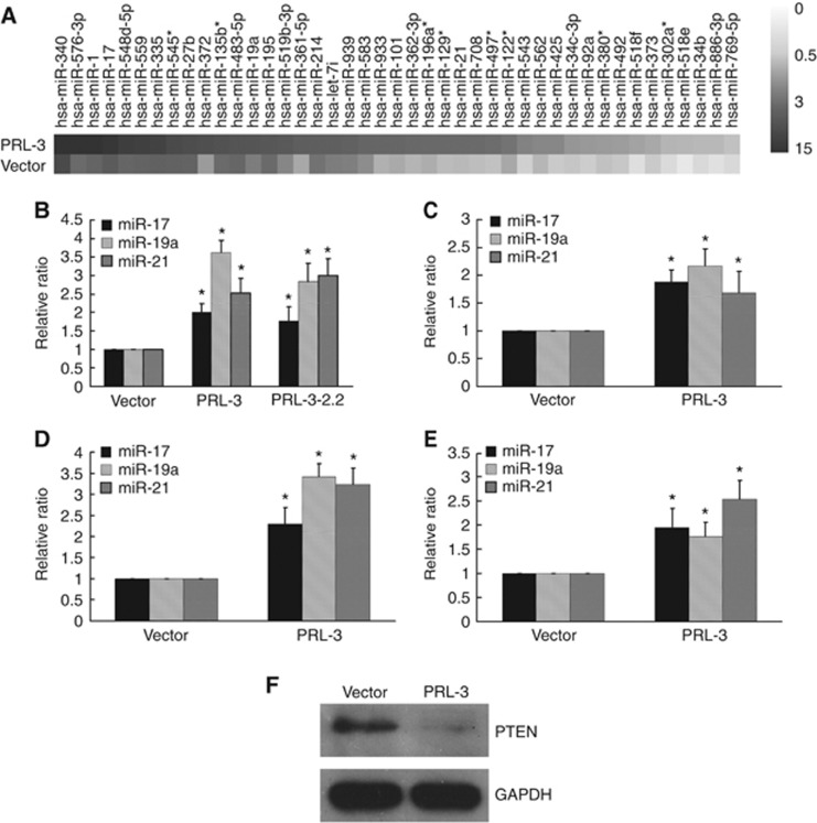
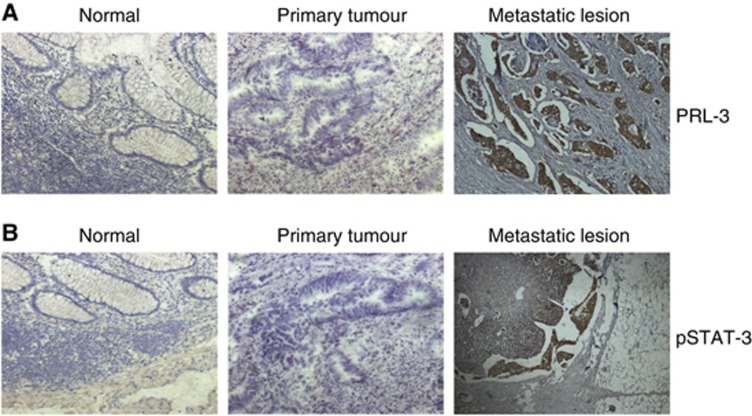
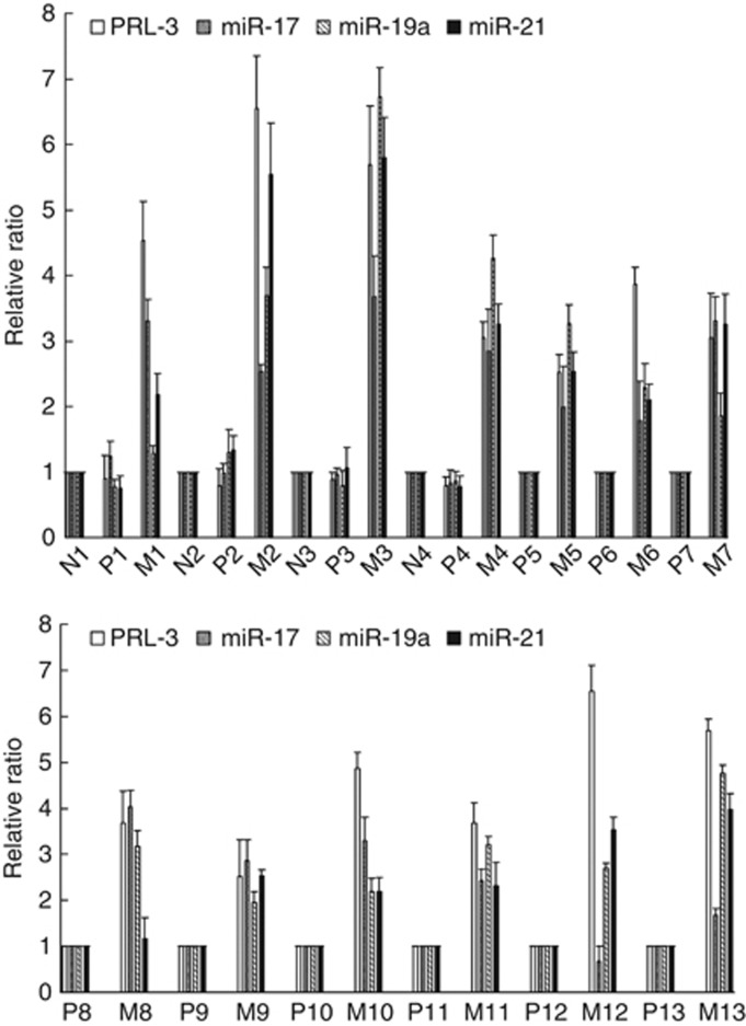
Methods: Microarray analysis and quantitative reverse-transcription polymerase chain reactions (qRT-PCR) were used to investigate the changes in miRNA expression due to the overexpression of PRL-3. Transwell chamber invasion assays, CCK-8 proliferation assays and RNA interference assays were used to explore the effects of PRL-3 on miR-21, miR-17 and miR-19a expression in colon cancer cells. Immunohistochemistry and qRT-PCR were performed in colon cancer tissues to evaluate the expression of PRL-3, signal transducer and activator of transcription 3 (STAT3), miR-21, miR-17 and miR-19a.

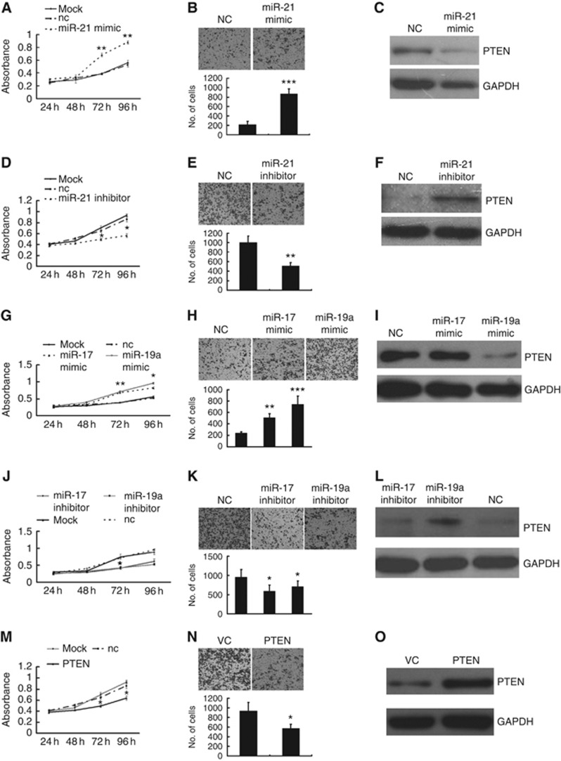
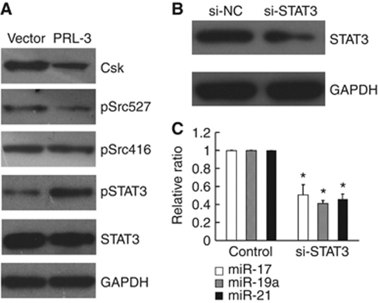
Results: Our study demonstrated that the overexpression of PRL-3 in colon cancer cells induced the expression of miR-21, miR-17 and miR-19a by activating STAT3. Subsequently, these microRNAs contributed to the increased proliferation and invasiveness of the colon cancer cells. Positive correlations between PRL-3 and these microRNAs were also observed in matched primary colon cancer tissues and metastatic lesions.

Conclusion: miR-21, miR-17 and miR-19a induced by PRL-3 contribute to the proliferation and invasion of colon cancer.


Figures

Figure 1
Figure 1
Overexpression of PRL-3 promoted the proliferation, migration and invasion of LoVo colon cancer cells. (A) qRT–PCR and (B) western blot analysis confirmed the expression of PRL-3 in the PRL-3 and PRL-3–2.2 cells; cells transfected with the empty vector were used as a negative control. (C) PRL-3 promoted the proliferation of LoVo cells in a colony formation assay and (D) a CCK-8 proliferation assay. (E) PRL-3 promoted the migration of PRL-3–2.2 cells in a wound-healing assay. (F) PRL-3 cells exhibited more invasive potential (versus the control) in a Transwell chamber invasion assay (*P<0.05, **P<0.01, ***P<0.001; Student’ t-test; n=3).
Figure 2
Figure 2
PRL-3 elevated the expression levels of miR-17, miR-19a, and miR-21 in CRC cells. (A) miRNA array analysis revealed that PRL-3 altered the expression of numerous microRNAs, including miR-17, miR-19a, and miR-21. LoVo cells that were (B) stably or (C) transiently transfected with PRL-3 exhibited elevated levels of miR-17, miR-19a and miR-21 expression, as determined by qRT–PCR. PRL-3 also significantly increased the expression of miR-17, miR-19a and miR-21 in both (D) SW480 and (E) caco2 cells that had been transiently transfected with the PRL-3 expression plasmid compared with cells transfected with the control vector. (F) PTEN, a suppressor of the PI3K pathway, was downregulated at the protein level in LoVo-PRL-3 cells (*P<0.05; Student’ t-test; n=3).
Figure 3
Figure 3
miR-21, miR-17, and miR-19a promoted the proliferation and migration of LoVo cells and directly targeted PTEN. (A and G) LoVo-VC cells overexpressing miR-21, miR-17, and miR-19a exhibited significantly enhanced proliferative capability, as determined by a CCK8 assay, and (B and H) an enhanced invasive ability, as determined by a Transwell assay. (D, E, J and K) However, miR-21, miR-17 and miR-19a inhibitors antagonised the proliferation and migration of LoVo-PRL-3 cells. (C and I) The PTEN protein, a known target of miR-21 and miR-17–92, was significantly downregulated by miR-21 and miR-19a mimics in LoVo-VC cells. (F and L) miR-21 and miR-19a inhibitors restored the expression of the PTEN protein in LoVo-PRL-3 cells. (O) Overexpressed PTEN in LoVo-PRL-3 cells. (M and N) PTEN antagonised the phenotype exerted by these miRNAs in proliferation and invasion assay in LoVo-PRL-3 cells (*P<0.01, **P<0.01; Student’ t-test; n=3).
Figure 4
Figure 4
PRL-3 activated STAT3, leading to the upregulation of miR-17, miR-19a and miR-21. (A) PRL-3 downregulated Csk and pSrc (Tyr527), which is an inactive form of Src, culminating in the phosphorylation of Tyr705 of STAT3; By contrast, total STAT3 and pSrc (Tyr416), which is an active form of Src, were not affected. (B) The knockdown of STAT3 with a specific siRNA in LoVo-PRL-3 cells lowered the expression of the STAT3 protein. (C) The expression levels of miR-17, miR-19a and miR-21 were significantly reduced upon STAT3 downregulation (*P<0.05; Student’ t-test; n=3).
Figure 5
Figure 5
PRL-3 and pSTAT3 were elevated in metastatic colon cancer lesions. (A) Strong PRL-3 expression can be observed in metastatic lesions, mainly in the cytoplasm, whereas no positive staining was observed in the paired primary tumour or normal colon tissues. (B) pSTAT3 was strongly expressed in metastatic lesions, no positive staining was observed in the corresponding primary tumour or normal colon tissue.
Figure 6
Figure 6
miR-17, miR-19a, and miR-21 were elevated upon the upregulation of PRL-3 in almost all of the metastatic tissues examined compared with the primary colon cancer samples (P-primary tumours; M-metastatic lesions; N-normal colon tissue).

References

    1. Asangani IA, Rasheed SA, Nikolova DA, Leupold JH, Colburn NH, Post S, Allgayer H (2008) MicroRNA-21 (miR-21) post-transcriptionally downregulates tumor suppressor Pdcd4 and stimulates invasion, intravasation and metastasis in colorectal cancer. Oncogene 27: 2128–2136 - PubMed
    1. Barbieri I, Pensa S, Pannellini T, Quaglino E, Maritano D, Demaria M, Voster A, Turkson J, Cavallo F, Watson CJ, Provero P, Musiani P, Poli V (2010) Constitutively active Stat3 enhances neu-mediated migration and metastasis in mammary tumors via upregulation of Cten. Cancer Res 70: 2558–2567 - PubMed
    1. Bardelli A, Saha S, Sager JA, Romans KE, Xin B, Markowitz SD, Lengauer C, Velculescu VE, Kinzler KW, Vogelstein B (2003) PRL-3 expression in metastatic cancers. Clin Cancer Res 9: 5607–5615 - PubMed
    1. Bollrath J, Greten FR (2009) IKK/NF-kappaB and STAT3 pathways: central signalling hubs in inflammation-mediated tumour promotion and metastasis. EMBO Rep 10: 1314–1319 - PMC - PubMed
    1. Brock M, Trenkmann M, Gay RE, Michel BA, Gay S, Fischler M, Ulrich S, Speich R, Huber LC (2009) Interleukin-6 modulates the expression of the bone morphogenic protein receptor type II through a novel STAT3-microRNA cluster 17/92 pathway. Circ Res 104: 1184–1191 - PubMed

Publication types

MeSH terms