. 2012 Sep 15;21(18):3993-4006.
doi: 10.1093/hmg/dds222. Epub 2012 Jun 7.

Functional evaluation of BRCA2 variants mapping to the PALB2-binding and C-terminal DNA-binding domains using a mouse ES cell-based assay

Affiliations

Functional evaluation of BRCA2 variants mapping to the PALB2-binding and C-terminal DNA-binding domains using a mouse ES cell-based assay

Kajal Biswas et al. Hum Mol Genet. .

Abstract

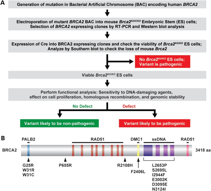
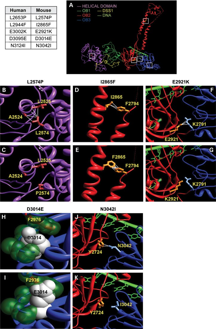
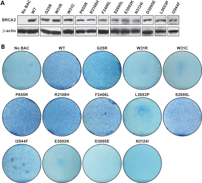
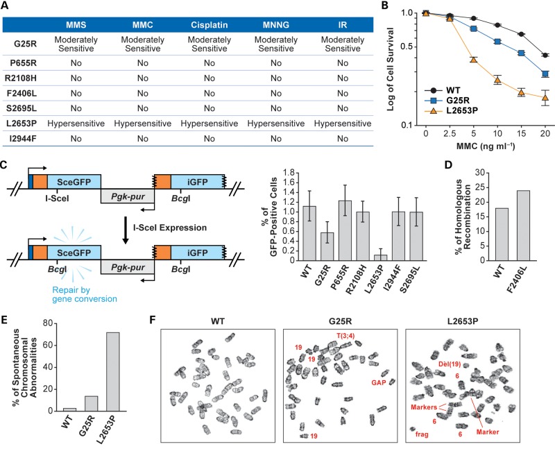
Single-nucleotide substitutions and small in-frame insertions or deletions identified in human breast cancer susceptibility genes BRCA1 and BRCA2 are frequently classified as variants of unknown clinical significance (VUS) due to the availability of very limited information about their functional consequences. Such variants can most reliably be classified as pathogenic or non-pathogenic based on the data of their co-segregation with breast cancer in affected families and/or their co-occurrence with a pathogenic mutation. Biological assays that examine the effect of variants on protein function can provide important information that can be used in conjunction with available familial data to determine the pathogenicity of VUS. In this report, we have used a previously described mouse embryonic stem (mES) cell-based functional assay to characterize eight BRCA2 VUS that affect highly conserved amino acid residues and map to the N-terminal PALB2-binding or the C-terminal DNA-binding domains. For several of these variants, very limited co-segregation information is available, making it difficult to determine their pathogenicity. Based on their ability to rescue the lethality of Brca2-deficient mES cells and their effect on sensitivity to DNA-damaging agents, homologous recombination and genomic integrity, we have classified these variants as pathogenic or non-pathogenic. In addition, we have used homology-based modeling as a predictive tool to assess the effect of some of these variants on the structural integrity of the C-terminal DNA-binding domain and also generated a knock-in mouse model to analyze the physiological significance of a residue reported to be essential for the interaction of BRCA2 with meiosis-specific recombinase, DMC1.


Figures

Figure 1.
Figure 1.
Scheme to examine human BRCA2 variants using the mES cell-based assay and the location of variants on the protein. (A) Schematic representation of the mES cell-based assay for functional analysis of BRCA2. bacterial artificial chromosome (BAC) DNA containing desired variants was first introduced into mES cells with one knockout allele (KO) and one conditional allele (CKO) of Brca2. After Cre-mediated deletion of a conditional copy of Brca2 depending on the impact of the variants, cells may or may not be viable. The viable ES cells can be functionally similar to WT or defective in some function of BRCA2, depending on the effect of variants. (B) Schematic diagram of the BRCA2 protein showing the different domains and position of the variants analyzed in this study. The BRCA2-interacting partners are shown above the corresponding region of BRCA2 required for their interaction. Below the location of the variants used in this study are shown.
Figure 2.
Figure 2.
Homology–based modeling of BRCA2 variants based on crystal structure of the mouse protein (A) The crystal structure of mouse C-terminal BRCA2 includes multiple domains: helical (magenta), OB1 (dark green), OB2 (red) and OB3 (blue). While the helical and OB1 domains interact with the co-factor DSS1, OB2 and OB3 domains interact with the ssDNA. The variants studied in this work are indicated by white squares. Human BRCA2 variants and corresponding residues in the mouse protein are indicated in the table on left. (B) L2574 forms hydrophobic contacts with residues A2524 and L2525, and together these residues form a core of the helical domain. (C) P2574 is not hydrophobic and loses these crucial contacts. (D) I2865 is present at the tower structure of OB2 and forms hydrophobic contacts with F2794, which could be crucial for tower structure. (E) Being hydrophobic, F2865 maintains these contacts. (F) At the interface between OB2 and OB3, E2921 forms several contacts and an electrostatic salt bridge with R2971. (G) E2921K mutation reverses the charge of the residue, introducing repulsive interactions and hence disrupting the crucial salt bridge and other contacts. (H) D3014 is placed on a beta-strand at the core of OB3. The side chain of D3014 is stacked up between the backbone of the beta-strand and the F2976 of an adjacent helix. (I) E3014 has a larger chain length and surface area, which clashes with the F2976. (J) N3042 is present at the beta-sheet interface between OB2 and OB3 and has two contacts across the interface with Y2724. (K) I3042 loses these contacts, which could disturb the proper packing arrangement of these domains. Except (H) and (I), the side chains of the helical domain are colored brown, OB2 are colored orange and OB3 are colored light blue. In (H) and (I), key atoms are shown as spheres with Van der Waals radii. Atoms of D3014 and E3014 are colored white, whereas the other key atoms are colored dark green.
Figure 3.
Figure 3.
Mouse ES cell-based assay for functional assay of BRCA2 variants: (A) western blot showing the expression of human BRCA2 variants in mES cells before removal of the conditional copy of Brca2. Antibody against c-myc epitope was used to detect the c-myc-tagged BRCA2. β-Actin was used as control. (B) Methylene blue staining of HATr colonies of ES cells expressing no BAC, WT or different variants of BRCA2.
Figure 4.
Figure 4.
Functional evaluation of BRCA2 variants: (A) sensitivity of the Brca2KO/KO ES cells expressing human BRCA2 variants to different DNA-damaging agents. No, no significant difference in the sensitivity compared with the Brca2KO/KO ES cells expressing WT human BRCA2. (B) Survival of ES cells expressing WT, BRCA2G25R or BRCA2L2653P exposed to MMC. P-values for BRCA2G25R is 0.0198 and for BRCA2L2653P is 0.0098 at 10 ng/ml MMC. (C) HR assay using direct repeats of mutated green fluorescent protein (DR-GFP) (37). Upon expression of I-SceI in the cells, a DSB is induced in the SceGFP construct. HR using the promoterless downstream iGFP as template generates an intact GFP protein and can be monitored by cellular green fluorescence. Right panel shows the percentages of GFP-positive cells after expressing I-SceI. (D) HR efficiency as measured by gene targeting at the Rosa26 locus (24). (E) Histogram showing the total number of spontaneous chromosomal abnormalities in cells expressing WT, G25R or L2653P BRCA2. Chromosomal abnormalities observed in WT: breaks/gaps 1%, fragments 2%; in G25R: breaks/gaps 5%, dicentric chromosomes 2%, fragments 4% and marker chromosomes 3%; in L2653P: breaks/gaps 21%, dicentric chromosomes 11%, fragments 17%, radial 2% and marker chromosomes 21%. A marker chromosome is an abnormal chromosome that is distinctive in appearance but not fully identified. Euploidy is not included in these abnormalities. (F) Panels showing representative metaphase spread of cells expressing WT, G25R and L2653P BRCA2. Arrows mark the chromosomal abnormalities; 19 refers to three copies of chromosome 19 in G25R and 6 refers to three copies of chromosome 6 in L2653P. To rule out the possibility of secondary mutations, each experiment was conducted using at least two independent clones.

References

    1. Fackenthal J.D., Olopade O.I. Breast cancer risk associated with BRCA1 and BRCA2 in diverse populations. Nat. Rev. Cancer. 2007;7:937–948. - PubMed
    1. Ford D., Easton D.F., Stratton M., Narod S., Goldgar D., Devilee P., Bishop D.T., Weber B., Lenoir G., Chang-Claude J., et al. Genetic heterogeneity and penetrance analysis of the BRCA1 and BRCA2 genes in breast cancer families. The Breast Cancer Linkage Consortium. Am. J. Hum. Genet. 1998;62:676–689. - PMC - PubMed
    1. Chen S., Parmigiani G. Meta-analysis of BRCA1 and BRCA2 penetrance. J. Clin. Oncol. 2007;25:1329–1333. - PMC - PubMed
    1. Rahman N., Stratton M.R. The genetics of breast cancer susceptibility. Annu. Rev. Genet. 1998;32:95–121. - PubMed
    1. Nathanson K.L., Wooster R., Weber B.L. Breast cancer genetics: what we know and what we need. Nat. Med. 2001;7:552–556. - PubMed

Publication types

MeSH terms