. 2012 Sep;33(9):1769-81.
doi: 10.1093/carcin/bgs200. Epub 2012 Jun 7.

A novel microtubule-modulating agent EM011 inhibits angiogenesis by repressing the HIF-1α axis and disrupting cell polarity and migration

Affiliations

A novel microtubule-modulating agent EM011 inhibits angiogenesis by repressing the HIF-1α axis and disrupting cell polarity and migration

Prasanthi Karna et al. Carcinogenesis. 2012 Sep.

Abstract

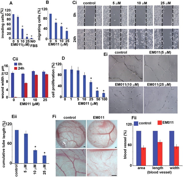
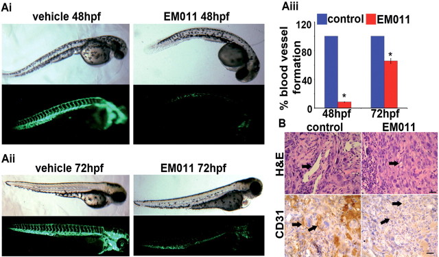
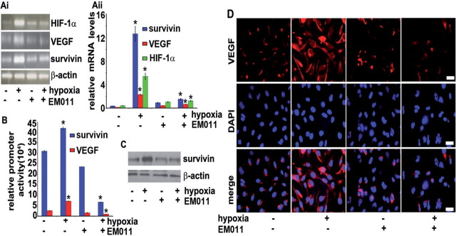
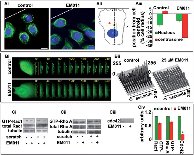
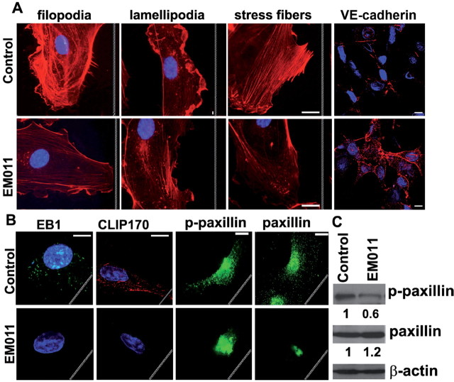
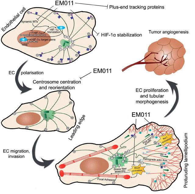
Endothelial tubular morphogenesis relies on an exquisite interplay of microtubule dynamics and actin remodeling to propel directed cell migration. Recently, the dynamicity and integrity of microtubules have been implicated in the trafficking and efficient translation of the mRNA for HIF-1α (hypoxia-inducible factor), the master regulator of tumor angiogenesis. Thus, microtubule-disrupting agents that perturb the HIF-1α axis and neovascularization cascade are attractive anticancer drug candidates. Here we show that EM011 (9-bromonoscapine), a microtubule-modulating agent, inhibits a spectrum of angiogenic events by interfering with endothelial cell invasion, migration and proliferation. Employing green-fluorescent transgenic zebrafish, we found that EM011 not only inhibited vasculogenesis but also disrupted preexisting vasculature. Mechanistically, EM011 caused proteasome-dependent, VHL-independent HIF-1α degradation and repressed expression of HIF-1α downstream targets, namely VEGF and survivin. Furthermore, EM011 inhibited membrane ruffling and impeded formation of filopodia, lamellipodia and stress fibers, which are critical for cell migration. These events were associated with a drug-mediated decrease in activation of Rho GTPases- RhoA, Cdc42 and Rac1, and correlated with a loss in the geometric precision of centrosome reorientation in the direction of movement. This is the first report to describe a previously unrecognized, antiangiogenic property of a noscapinoid, EM011, and provides evidence for novel anticancer strategies recruited by microtubule-modulating drugs.


Figures

Fig. 1.
Fig. 1.
EM011 inhibited the invasion and migration of endothelial cells in response to a chemotactic gradient. HUVECs were harvested and seeded onto 8 μm transwell-inserts (upper-chamber) coated with Matrigel in the presence of EM011 for 24h for the invasion assay. At the end of incubation times, cells in the upper chamber were removed with cotton swabs and cells that traversed the Matrigel to the lower surface of the insert were fixed with 10% formalin, stained with crystal violet, and counted under a light microscope. (A) Quantitation of the cells that invaded to the lower surface of the insert was carried out using three random fields/insert. The average number of invasive cells under control conditions (0.1% DMSO) was determined from three random fields and designated as the ‘100%’ value. For each treatment, the number of invasive cells in presence of EM011 was expressed as cells/field (mean ± SD, n = 3), quantitated as a percentage of the ‘100%’ value for the control. Migratory response was evaluated by coating the cell culture inserts underside with Matrigel and the cells were seeded next day onto the inserts in the upper chamber in the presence or absence of drug. The effect of drug on endothelial cell migration was observed by their inclusion in the lower chamber using crystal violet staining as described for the cell invasion assay. (B) Quantitation of cells that migrated to the lower surface of the insert was performed by counting cells from three random fields/insert. The average number of cells that had migrated under control conditions (0.1% DMSO) was determined from three random fields and designated as the ‘100%’ value. For each treatment, the number of cells that had migrated in presence of EM011 was expressed as cells/field (mean ± SD, n = 3), quantitated as a percentage of the ‘100%’ value for the control. Data are expressed as cells/field (mean ± SD) as a percentage of control. EM011 inhibited endothelial cell migration in a chemokinetic assay. (Ci) Photomicrographs (×10) showing confluent HUVECs that were mechanically scraped with a pipette tip, and the migration of HUVECs into the scraped area after 24h when treated with DMSO (control) or varying EM011 doses (5, 10, 25 µM). Dotted line indicates the denuded area occupied by the initial scraped area. (Cii) Quantitation of the extent of wound closure upon various treatments as measured by the width in µm of the denuded space. (D) shows the dose-dependent inhibition of HUVEC proliferation upon EM011 treatment using the sulforhodamine B assay. (Ei) EM011 inhibited in vitro endothelial tubule formation. The spontaneous formation of capillary-like structures by HUVECs on Matrigel was used to assess the angiogenic potential. HUVECs were seeded onto Matrigel and 1h later, EM011 was added for 16h. Photomicrographs show that vehicle-treated controls migrated to form connected tubular networks, whereas EM011 significantly attenuated tubular morphogenesis and network formation. (Eii) Inhibition of endothelial tubule formation upon a 16h EM011 treatment was quantified from photographs by measuring the cumulative tube length contained in two random fields from each well under a phase-contrast microscope. Data are expressed as a percentage of the cumulative tube length in vehicle-treated cultures (mean ± SD). (Fi) Ex vivo antiangiogenic activity of EM011 in a CAM assay. Normal angiogenesis observed in CAM development in controls (Fi, left panel) versus decreased vascularization in the development of the CAM (Fi, right panel) when treated with 25 µM EM011. Scale bar = 5 µm. (Fii) illustrates a marked decrease in blood vessel area, length and width upon treatment with 25 μM EM011 compared with control vessels over a 24h period.
Fig. 2.
Fig. 2.
(Ai) EM011 inhibits angiogenic vessel growth in zebrafish embryos. Upper-panel (bright field) and lower-panel (fluorescent) depict images of 24 hpf zebrafish embryos treated for 24h with 1% DMSO (control) or 25 µM EM011. Significant inhibition of the outgrowth of intersegmental vessels indicates strong antiangiogenic effects of EM011. (Aii) EM011 disrupts preexisiting vasculature. 48 hpf embryos were treated with the drug for 24h. These embryos were imaged at 72 hpf when the vasculature is normally stabilized. The absence of intersegmental vessels and faint presence of the vasculogenic vessels show that EM011 significantly collapsed preexisting vasculature and promoted instability of these vessels. (Aiii) Quantification of intersegmental vessels upon 24h drug treatment of 24 hpf and 48 hpf embryos (*P < 0.05 versus control). Intersegmental vessels were first quantitated from 20 zebrafish embryos grown under control conditions and this value was designated at the ‘100%’ value. The number of intersegmental vessels were then determined for 20 embryos grown under each treatment condition, and each of these values was quantitated as a percentage of the ‘100%’ value for the control. (B) EM011 inhibited in vivo angiogenesis in a Matrigel plug assay. Photomicrographs show the effect of 7 day oral EM011 feeding on angiogenic response to FGF-2 supplemented PC-3 cells subcutaneously implanted as Matrigel plugs in mice. Blood vessel formation was examined by H&E and CD31 immunostaining of excised Matrigel plugs from mice that received vehicle or EM011 daily at a dose level of 300mg/kg by oral gavage. In the control images, arrows (in H&E micrographs) indicate subcutaneous Matrigel implants that demonstrate increased angiogenesis, seen as well-developed, endothelial cell–lined vascular spaces. This contrasts with the poorly developed vascular channels in the EM011 treated group. Arrows in the CD31 immunostained micrographs highlight the endothelial cells, with increased expression observed in the control group suggesting increased angiogenesis. Scale bar = 20 µm.
Fig. 3.
Fig. 3.
EM011 decreased HIF-1α protein levels in a VHL-independent pathway. PC-3 cells were exposed to hypoxia in the presence or absence of 25 µM EM011 for (A) various time points under hypoxia, or (B) 24h under normoxia or hypoxia. (C) Immunofluorescence staining of HIF-1α in normoxic or hypoxic samples of PC-3 cells treated with 25 µM EM011 for 24h. Scale bar = 20 µm. (D) HIF-1α–dependent HRE-luc activity determined in PC-3 cells co-transfected for 6h with 6xHRE luc and pRL-SV40 Renilla luciferase plasmids followed by a 24h treatment with 0, 1, 10, 25 or 50 µM EM011 under hypoxia (*P < 0.05 versus control). (E) RCC4 cells were treated with 25 µM EM011 for 24h under normoxia and cell lysates were immunoblotted for HIF-1α protein expression. (F) Proteasome inhibition with 5 µM MG132 abrogates EM011-mediated HIF-1α degradation. PC-3 cells were co-treated with MG132 and EM011 treatment for 16h in the presence or absence of hypoxia. Cell lysates were processed for immunoblotting for HIF-1α expression.
Fig. 4.
Fig. 4.
EM011 induces transcriptional repression of HIF-1α and its downstream target genes, VEGF and survivin. (Ai) EM011 decreases mRNA levels of HIF-1α, survivin and VEGF. PC-3 cells were treated with 25 µM drug in the presence or absence of hypoxia for 24h and total RNA was extracted followed by cDNA synthesis, which was quantified by real-time RT-PCR using specific primers. (Aii) Quantitation of relative mRNA levels. (B) EM011 inhibits HIF-1α transcriptional activity as seen by decreased VEGF or survivin promoter activity in a dual-luciferase promoter activity assay. (C) Immunoblot showing survivin levels under the noted treatment regimes. (D) Confocal immunofluorescence micrographs showing VEGF staining in control and EM011-treated cells under hypoxia or normoxia. Scale bar = 20 µm (*P < 0.05 versus control).
Fig. 5.
Fig. 5.
(Ai) EM011 impairs centrosome repositioning and disrupts directed migration. Confluent HUVEC monolayers were mechanically scraped to stimulate unidirectional migration, and the position of the centrosome in relation to the nucleus was visualized after 24h in the first row of cells adjacent to the open edge of the monolayer by immunofluorescent staining of α-tubulin (in green) for microtubules, γ-tubulin (in red) for centrosomes and DAPI (blue) for nuclei. White broken line indicates the LE. Scale bar = 20 µm. (Aii) An illustration of centration analysis that was performed by comparing the location of the nuclear and centrosomal centroids with the cell centroid. EM011-treated cells displayed a disruption in backward nuclear movement with respect to controls, indicating a perturbation in the migratory process. (Aiii) Graphical representation of the position from the cell centroid clearly displays a failure of rearward nuclear movement upon EM011 treatment. (Bi) HUVECs were transfected with pEGFPC1 followed by a scratch in the presence and absence of EM011. GFP fluorescence at the LE of cells was recorded at 40 s intervals using the TCS SP5 confocal microscope (Leica), equipped with a live-cell imaging workstation and the LASAF software. Rectangular regions were selected as indicated to analyze membrane ruffle dynamics. (Bii) Membrane ruffle dynamics was presented as three-dimensional surface plots. The y-axes of the plots denote the extent of protrusion of the region of the membrane ruffle delineated by the rectangle. (Ci–iii) HUVECs treated with vehicle or EM011 were scratched, and the level of activated (GTP-bound) Rac1, RhoA and Cdc42 were assayed by immunoblot analysis of the GST-PBD/GST-RBD pull-down preparation with anti-Rac1, anti-Cdc42 and anti-RhoA antibodies. The levels of total Rac1, RhoA and tubulin were examined by immunoblot analysis of cell lysates. (Civ) Relative activity of Rac1, RhoA and Cdc42 was measured by densitometric analysis of the blots. Data are the mean and standard error from three experiments (*P < 0.05 versus control).
Fig. 6.
Fig. 6.
EM011 affects actin structures and adherens junctions involved in endothelial cell migration. (A) HUVECs scratched in the absence or presence of EM011 were stained for F-actin to visualize filopodia (filamentous membrane projections with long parallel actin filaments arranged in tight bundles), lamellipodia (cytoplasmic protrusions that contain a thick cortical network of actin filaments) and stress fibers (bundles of actin filaments anchored at FAs required for the traction of the rear of the cells toward the LE during migration). Endothelial cells were stained with VE-cadherin (red) and nuclei (blue). Interestingly, the VE-cadherin levels increased and appeared to get redistributed to the cellular periphery, particularly evident at sites of cell–cell contacts. Scale bar = 20 µm. (B) Immunofluorescence micrographs showing localization of EB1, CLIP170, paxillin and p-paxillin in HUVECs that were scratched in the presence or absence of EM011. Scale bar = 20 µm. White jagged lines indicate the LE. (C) EM011 diminished tyrosine phosphorylation of paxillin, but not total paxillin expression, in HUVECs. HUVECs were scratched in the presence or absence of 10 µM EM011 for 2h. The cell lysates were subjected to immunoblotting with anti-phospho-paxillin and anti-paxillin. Values are from densitometric results of three separate experiments. *P < 0.05, EM011 treated versus control cells.
Fig. 7.
Fig. 7.
Schematic illustration of a working model to demonstrate that EM011 treatment disrupts various aspects of HIF-1α–dependent signaling and impairs cell polarization, migration and tumor angiogenesis. EM011 treatment decreases the transcription of HIF-1α by mechanisms yet to be identified. By attenuating microtubule dynamicity, EM011 also inhibits transport and translation of HIF-1α mRNA and induces proteasome-dependent degradation of HIF-1α. The net decrease in HIF-1α protein levels causes a reduction in expression of HIF-1α–responsive genes, including VEGF and survivin, that are essential for tumor angiogenesis. Reduced microtubule dynamicity also causes diminished binding of plus-end tracking proteins such as EB1 and CLIP-170 to microtubule plus-ends. EM011 inhibits the processes of centrosome centration and reorientation toward the direction of migration, which are crucial to the cell’s ability to perceive and alter the geometry of its actin and microtubule cytoskeletons in order to generate asymmetry between the cell front and rear for locomotion. Migrating cells develop a broad, flat lamellipodium by nucleating actin polymerization at the LE to produce a highly cross-linked meshwork of actin filaments (shown with red lines) whose growing ends face the cell front. Outgrowth of these filaments pushes the LE forward and generates retrograde flow of actin (shown by blue arrows) toward the cell center. Microtubules undergo net growth near the LE (depicted with green arrows) and activate the Rac1 GTPase which is responsible for lamellipodial protrusion and the formation of short-lived, small focal complexes at the lamellipodium base. Microtubules subjected to actin retrograde flow tend to bend and break in the cell body creating depolymerizing microtubule minus-ends and leading to RhoA activation in the cell body. EM011 hinders RhoA activation which normally drives assembly of contractile stress fibers and larger FAs. FAs are targeted by EB1- and CLIP-170–bound dynamic microtubule plus-ends, leading to FA disassembly and cell motility––a process also inhibited by EM011. Thus, EM011 drastically perturbs several crucial steps in endothelial cell migration and eventually, tumor angiogenesis.

Similar articles

Cited by

References

    1. Wittmann T, et al. (2001). Cell motility: can Rho GTPases and microtubules point the way? J. Cell Sci. 114 3795–3803 - PubMed
    1. Lauffenburger D.A., et al. (1996). Cell migration: a physically integrated molecular process Cell 84 359–369 - PubMed
    1. Waterman-Storer C.M., et al. (1999). Positive feedback interactions between microtubule and actin dynamics during cell motility Curr. Opin. Cell Biol. 11 61–67 - PubMed
    1. Small J.V., et al. (2003). Microtubules meet substrate adhesions to arrange cell polarity Curr. Opin. Cell Biol. 15 40–47 - PubMed
    1. Mabjeesh N.J., et al. (2003). 2ME2 inhibits tumor growth and angiogenesis by disrupting microtubules and dysregulating HIF Cancer Cell 3 363–375 - PubMed

Publication types