. 2012 May 20;486(7401):135-9.
doi: 10.1038/nature11077.

Crystal structure of a voltage-gated sodium channel in two potentially inactivated states

Affiliations

Crystal structure of a voltage-gated sodium channel in two potentially inactivated states

Jian Payandeh et al. Nature. .

Abstract

In excitable cells, voltage-gated sodium (Na(V)) channels activate to initiate action potentials and then undergo fast and slow inactivation processes that terminate their ionic conductance. Inactivation is a hallmark of Na(V) channel function and is critical for control of membrane excitability, but the structural basis for this process has remained elusive. Here we report crystallographic snapshots of the wild-type Na(V)Ab channel from Arcobacter butzleri captured in two potentially inactivated states at 3.2 Å resolution. Compared to previous structures of Na(V)Ab channels with cysteine mutations in the pore-lining S6 helices (ref. 4), the S6 helices and the intracellular activation gate have undergone significant rearrangements: one pair of S6 helices has collapsed towards the central pore axis and the other S6 pair has moved outward to produce a striking dimer-of-dimers configuration. An increase in global structural asymmetry is observed throughout our wild-type Na(V)Ab models, reshaping the ion selectivity filter at the extracellular end of the pore, the central cavity and its residues that are analogous to the mammalian drug receptor site, and the lateral pore fenestrations. The voltage-sensing domains have also shifted around the perimeter of the pore module in wild-type Na(V)Ab, compared to the mutant channel, and local structural changes identify a conserved interaction network that connects distant molecular determinants involved in Na(V) channel gating and inactivation. These potential inactivated-state structures provide new insights into Na(V) channel gating and novel avenues to drug development and therapy for a range of debilitating Na(V) channelopathies.


Figures

Figure 1
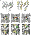
Figure 1. Structure and function of NavAb
a, Use-dependent development of slow inactivation. Depolarizations from a holding potential of −180 mV to −40 mV, 7 ms in duration, were applied at 0.2 Hz (circles) or once per min (triangles), and the peak current elicited by each pulse was measured. Currents were normalized to the peak inward current during the first pulse. b, Main structural elements of NavAb. The nearest VSD and pore domain are removed for clarity. c, Packing arrangement in the WT NavAb crystals. Four crystallographically independent channel subunits are colored: purple (chain A), yellow (chain B), cyan (chain C) and pink (chain D). d–e, Left, red dashed lines indicate the Cα location of D219 (the last S6 residue modeled in WT-AB), where the S6 helices are shown as cylinders. WT-chain A, purple; WT- chain B, yellow. Middle and right, space filling models in expanded boxes highlight Asn211 and the S6 interaction site of WT-chain A with Leu123 on the S4–S5 linker.
Figure 2
Figure 2. Structural changes in the selectivity filter of NavAb
a, Stick representation of the selectivity filter with a 2Fo–Fc map calculated at 3.2 Å resolution (grey mesh) contoured at 1.5 σ for NavAb-I217C and 1.0 σ for NavAb-AB and NavAb-CD. Symmetry-related subunits in WT-AB and WT-CD are colored white (Chains A and D) and cyan (Chains B and C), respectively. b, Hydrogen bonds (< 3.5 Å) supporting the selectivity, as discussed in the main text, are shown as dotted lines. c, Crystallographic b-factors are colored from low to high (blue to red) along the P-helix, selectivity filter, and P2-helix regions.
Figure 3
Figure 3. Conformational changes in the pore module of NavAb
a, Highly conserved residues in the PM of NaV channels are shown in stick and mesh representation. Mutations of the Leu170 and Ile202 side-chain equivalents (green and pink, respectively) in NaV1.4 alter slow-inactivation in NaV1.4. Asn211 (S6) is shown for reference. b, Superposition of the NavAb-I217C (grey) and NavAb-AB (yellow) PMs highlights a structurally-coupled, evolutionarily-conserved set of amino acid residues that communicate between the intracellular activation gate (S6), the central cavity (S5 and S6), and the selectivity filter (P- and P2-helices), and the selectivity filter. c, A view through the PM sectioned below the selectivity filter illustrates the lateral pore fenestrations, hydrophobic access to the central cavity, and structural asymmetry in the NavAb-AB and NavAb-CD pore domains. Phe203 side-chains are yellow sticks. NavAb residues implicated in drug binding in vertebrate NaV channels are colored: Thr206 (blue), Met209 (green), and Val213 (orange). d–e, Viewed from the plane of the lipid bilayer, the pore fenestrations provide dynamic access to the central cavity from the hydrophobic membrane phase. Phe203 side-chains are shown as stick and mesh representations (yellow). The yellow labels d and e on the structures in panel c show where the fenestrations illustrated in panels d and e are located in the complete structure. Note the positions of Phe203 illustrated in yellow in panels c-e for orientation.
Figure 4
Figure 4. Structure and coupling of the VSD in NavAb
a, Superposition of the S4 segment backbone from NavAb-I217C with WT-AB and WT-CD models (white, yellow, and salmon, respectively) shows high structural similarity. b, Crystallographic b-factors colored from low to high (blue to red) along one WT-AB subunit highlight the mobility of the S1–S3 segments relative to the S4 segment. c, The distribution of b-factors further suggests a structural coupling between the S4 segment, the S4–S5 linker, and the S5 segment. c, Superposition of the pore domains from NavAb-I217C (gray), WT-AB (yellow), and WT-CD (red) demonstrates a major hinge point at the base of the S5 segment. d, Selectivity filter-based superposition of the NavAb-AB and NavAb-CD channels illustrates a rolling motion of the VSDs around the PM during inactivation gating as viewed from the extracellular side of the membrane. Displacements of the VSDs measure up to ~5 Å. e, View from the plane of the membrane highlights the relative vertical and horizontal displacements of the WT NavAb VSDs during inactivation gating. Chain B, yellow; Chain C, red. f, Similar view of Chain A, yellow; Chain D, red. The labels on the structures in panel d show where the fenestrations illustrated in panels e and f are located in the complete structure.

References

    1. Hodgkin AL, Huxley AF. A quantitative description of membrane current and its application to conduction and excitation in nerve. J Physiol. 1952;117:500–544. - PMC - PubMed
    1. Rudy B. Slow inactivation of the sodium conductance in squid giant axons. Pronase resistance. J Physiol(Lond) 1978;283:1–21. - PMC - PubMed
    1. Vilin YY, Ruben PC. Slow inactivation in voltage-gated sodium channels: molecular substrates and contributions to channelopathies. Cell Biochem Biophys. 2001;35:171–190. - PubMed
    1. Payandeh J, Scheuer T, Zheng N, Catterall WA. The crystal structure of a voltage-gated sodium channel. Nature. 2011;475:353–358. - PMC - PubMed
    1. Long SB, Tao X, Campbell EB, MacKinnon R. Atomic structure of a voltage-dependent K+ channel in a lipid membrane-like environment. Nature. 2007;450:376–382. - PubMed

Publication types

Substances

Associated data