

dSarm/Sarm1 is required for activation of an injury-induced axon death pathway
- PMID: 22678360
- PMCID: PMC5225956
- DOI: 10.1126/science.1223899
dSarm/Sarm1 is required for activation of an injury-induced axon death pathway
Abstract
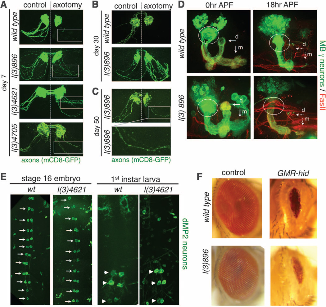
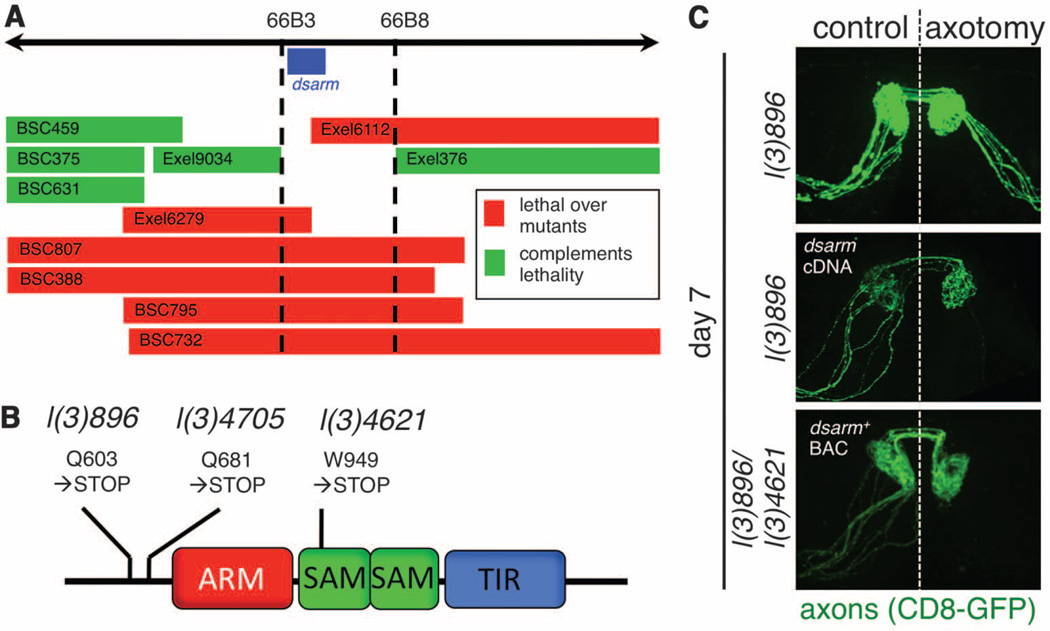
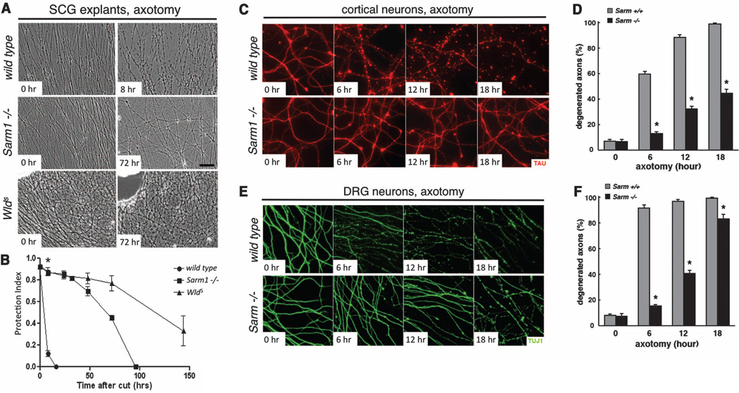
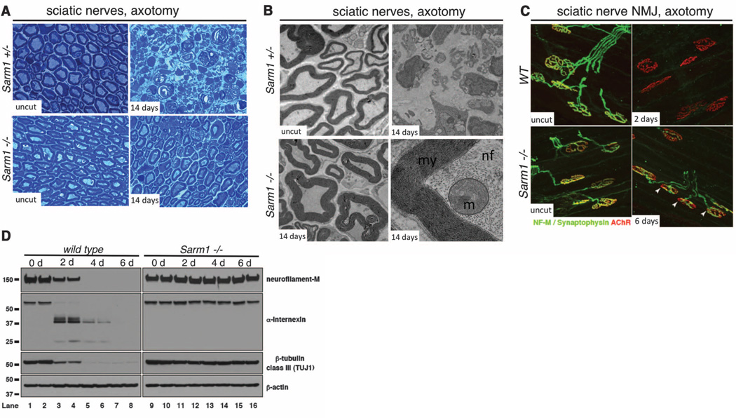
Axonal and synaptic degeneration is a hallmark of peripheral neuropathy, brain injury, and neurodegenerative disease. Axonal degeneration has been proposed to be mediated by an active autodestruction program, akin to apoptotic cell death; however, loss-of-function mutations capable of potently blocking axon self-destruction have not been described. Here, we show that loss of the Drosophila Toll receptor adaptor dSarm (sterile α/Armadillo/Toll-Interleukin receptor homology domain protein) cell-autonomously suppresses Wallerian degeneration for weeks after axotomy. Severed mouse Sarm1 null axons exhibit remarkable long-term survival both in vivo and in vitro, indicating that Sarm1 prodegenerative signaling is conserved in mammals. Our results provide direct evidence that axons actively promote their own destruction after injury and identify dSarm/Sarm1 as a member of an ancient axon death signaling pathway.
Figures
Comment in
-
Axon degeneration: A new pathway emerges.Nat Rev Neurosci. 2012 Jun 27;13(8):516. doi: 10.1038/nrn3294. Nat Rev Neurosci. 2012. PMID: 22735695 No abstract available.
-
Neuroscience. dSarm-ing axon degeneration.Science. 2012 Jul 27;337(6093):418-9. doi: 10.1126/science.1226150. Science. 2012. PMID: 22837513 No abstract available.
References
-
- Waller A. Philos. Trans. R. Soc. London Ser. B Biol. Sci. 1850;140:423.
-
- Lunn ER, Perry VH, Brown MC, Rosen H, Gordon S. Eur. J. Neurosci. 1989;1:27. - PubMed
-
- Glass JD, Brushart TM, George EB, Griffin JW. J Neurocytol. 1993;22:311. - PubMed
-
- Raff MC, Whitmore AV, Finn JT. Science. 2002;296:868. - PubMed
-
- Coleman MP, Perry VH. Trends Neurosci. 2002;25:532. - PubMed
Publication types
MeSH terms
Substances
Grants and funding
- R37 NS053538/NS/NINDS NIH HHS/United States
- HHMI/Howard Hughes Medical Institute/United States
- R01NS072248/NS/NINDS NIH HHS/United States
- R01 NS059991/NS/NINDS NIH HHS/United States
- 5R01-NS050557-05/NS/NINDS NIH HHS/United States
- R01 AI030165/AI/NIAID NIH HHS/United States
- U54NS065712/NS/NINDS NIH HHS/United States
- RC2-NS070-342/NS/NINDS NIH HHS/United States
- R01 NS050557/NS/NINDS NIH HHS/United States
- AI030165/AI/NIAID NIH HHS/United States
- BBS/E/B/000C0417/BB_/Biotechnology and Biological Sciences Research Council/United Kingdom
- R01 NS072248/NS/NINDS NIH HHS/United States
- U54 NS065712/NS/NINDS NIH HHS/United States
- RC2 NS070342/NS/NINDS NIH HHS/United States
- R01NS059991/NS/NINDS NIH HHS/United States
- BBS/E/B/0000C200/BB_/Biotechnology and Biological Sciences Research Council/United Kingdom
LinkOut - more resources
Full Text Sources
Other Literature Sources
Molecular Biology Databases
