

Mechanisms of ATP release and signalling in the blood vessel wall
- PMID: 22678409
- PMCID: PMC3400358
- DOI: 10.1093/cvr/cvs187
Mechanisms of ATP release and signalling in the blood vessel wall
Abstract
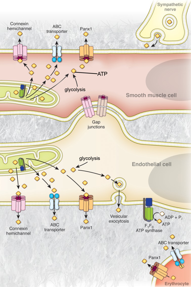
The nucleotide adenosine 5'-triphosphate (ATP) has classically been considered the cell's primary energy currency. Importantly, a novel role for ATP as an extracellular autocrine and/or paracrine signalling molecule has evolved over the past century and extensive work has been conducted to characterize the ATP-sensitive purinergic receptors expressed on almost all cell types in the body. Extracellular ATP elicits potent effects on vascular cells to regulate blood vessel tone but can also be involved in vascular pathologies such as atherosclerosis. While the effects of purinergic signalling in the vasculature have been well documented, the mechanism(s) mediating the regulated release of ATP from cells in the blood vessel wall and circulation are now a key target of investigation. The aim of this review is to examine the current proposed mechanisms of ATP release from vascular cells, with a special emphasis on the transporters and channels involved in ATP release from vascular smooth muscle cells, endothelial cells, circulating red blood cells, and perivascular sympathetic nerves, including vesicular exocytosis, plasma membrane F(1)/F(0)-ATP synthase, ATP-binding cassette (ABC) transporters, connexin hemichannels, and pannexin channels.
Figures
References
-
- Gribble FM, Loussouarn G, Tucker SJ, Zhao C, Nichols CG, Ashcroft FM. A novel method for measurement of submembrane ATP concentration. J Biol Chem. 2000;275:30046–30049. - PubMed
-
- Burnstock G. Neural nomenclature. Nature. 1971;229:282–283. - PubMed
-
- Lazarowski ER, Sesma JI, Seminario-Vidal L, Kreda SM. Molecular mechanisms of purine and pyrimidine nucleotide release. Adv Pharmacol. 2011;61:221–261. - PubMed
Publication types
MeSH terms
Substances
Grants and funding
LinkOut - more resources
Full Text Sources
Other Literature Sources
Miscellaneous
