

Review
doi: 10.1007/s10911-012-9258-0.
Epub 2012 Jun 8.
Determining mammosphere-forming potential: application of the limiting dilution analysis
Affiliations
- PMID: 22678420
- PMCID: PMC3428520
- DOI: 10.1007/s10911-012-9258-0
Item in Clipboard
Review
Determining mammosphere-forming potential: application of the limiting dilution analysis
J Mammary Gland Biol Neoplasia.
2012 Jun.
Abstract
Originally adapted from the neurosphere assay, the nonadherent mammosphere assay has been utilized to assess early progenitor/stem cell frequency in a given population of mammary epithelial cells. This method has also been used to measure the frequency of tumorsphere initiating cells in both primary mammary tumors as well as in tumor cell lines. Although, the mammosphere assay has been used extensively in the mammary gland field, a standard method of quantifying and analyzing sphere growth in this assay has remained undefined. Here, we discuss the use and benefit of using a limiting dilution analysis to quantify sphere-forming frequency in primary mammary epithelial cells grown in nonadherent conditions.
Figures
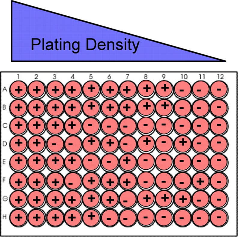
Schematic showing strategy for limiting dilution plating of dissociated epithelial cells in 96 well plate for SLDA. The “+” refers to a well containing one or more spheres and a “–” refers to a well without any spheres. The figure shows the cells being plated in a limiting dilution fashion across a 96-well plate. The amount of wells negative for sphere formation will increase as the plating density decreases as depicted
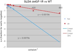
SLDA comparing stem cell frequency in tertiary spheres between dnhIGF-1R and FVB (WT) primary mammary epithelial cells grown in PRO-N media in 96 well ultra-low adherent plates (Corning). Plating density ranges from 1000 to 0.00012 cells/well, which was converted to cells/cm2 in this graph. Tertiary sphere forming frequency for dn-IGF-1R is 1 in 526 cells versus 1 in 127 cells for the wild type control. PRO-N media contained EGF/bFGF (20 ng/ml), bovine Insulin 25 μg/ml (Sigma), d-Biotin 10 ng/ml (Sigma), Progesterone 20 nM (Sigma), Putrescine 100 μM (Sigma), Selenium 5 ng/ml (Sigma), Apo-transferrin 50 μg/ml (Sigma), Gentamycin 50 μg/ml (Invitrogen-Gibco), Hydrocortisone 0.5 μg/ml (Sigma). Linear (fvb) and Linear (dn) indicate best fit line for linear regression analysis
Similar articles
-
A detailed mammosphere assay protocol for the quantification of breast stem cell activity.J Mammary Gland Biol Neoplasia. 2012 Jun;17(2):111-7. doi: 10.1007/s10911-012-9255-3. Epub 2012 Jun 5. J Mammary Gland Biol Neoplasia. 2012. PMID: 22665270 Review.
-
Assays for functionally defined normal and malignant mammary stem cells.Adv Cancer Res. 2019;141:129-174. doi: 10.1016/bs.acr.2018.12.004. Epub 2019 Jan 17. Adv Cancer Res. 2019. PMID: 30691682 Review.
-
Role of epithelial stem/progenitor cells in mammary cancer.Gene Expr. 2011;15(3):133-40. doi: 10.3727/105221611x13176664479368. Gene Expr. 2011. PMID: 22268295 Free PMC article. Review.
-
Introduction of SV40ER and hTERT into mammospheres generates breast cancer cells with stem cell properties.Oncogene. 2012 Apr 12;31(15):1896-909. doi: 10.1038/onc.2011.378. Epub 2011 Aug 29. Oncogene. 2012. PMID: 21874052
-
NOTCH1 inhibition in vivo results in mammary tumor regression and reduced mammary tumorsphere-forming activity in vitro.Breast Cancer Res. 2012 Sep 19;14(5):R126. doi: 10.1186/bcr3321. Breast Cancer Res. 2012. PMID: 22992387 Free PMC article.
Cited by
-
Monoamine oxidase-A activity is required for clonal tumorsphere formation by human breast tumor cells.Cell Mol Biol Lett. 2019 Nov 12;24:59. doi: 10.1186/s11658-019-0183-8. eCollection 2019. Cell Mol Biol Lett. 2019. PMID: 31754354 Free PMC article.
-
Early Dietary Exposures Epigenetically Program Mammary Cancer Susceptibility through Igf1-Mediated Expansion of the Mammary Stem Cell Compartment.Cells. 2022 Aug 17;11(16):2558. doi: 10.3390/cells11162558. Cells. 2022. PMID: 36010633 Free PMC article.
-
Organ-specific isogenic metastatic breast cancer cell lines exhibit distinct Raman spectral signatures and metabolomes.Oncotarget. 2017 Mar 21;8(12):20266-20287. doi: 10.18632/oncotarget.14865. Oncotarget. 2017. PMID: 28145887 Free PMC article.
-
TSSC3 represses self-renewal of osteosarcoma stem cells and Nanog expression by inhibiting the Src/Akt pathway.Oncotarget. 2017 Aug 24;8(49):85628-85641. doi: 10.18632/oncotarget.20429. eCollection 2017 Oct 17. Oncotarget. 2017. PMID: 29156746 Free PMC article.
-
A miRNA-Mediated Approach to Dissect the Complexity of Tumor-Initiating Cell Function and Identify miRNA-Targeting Drugs.Stem Cell Reports. 2019 Jan 8;12(1):122-134. doi: 10.1016/j.stemcr.2018.12.002. Stem Cell Reports. 2019. PMID: 30629937 Free PMC article.
References
Publication types
MeSH terms
Grants and funding
LinkOut - more resources
Full Text Sources
Other Literature Sources
Medical
