Review
. 2012 Aug;46(1):151-60.
doi: 10.1007/s12035-012-8281-x. Epub 2012 Jun 8.

Alzheimer's disease, oestrogen and mitochondria: an ambiguous relationship

Affiliations
Review

Alzheimer's disease, oestrogen and mitochondria: an ambiguous relationship

Amandine Grimm et al. Mol Neurobiol. 2012 Aug.

Abstract

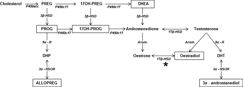
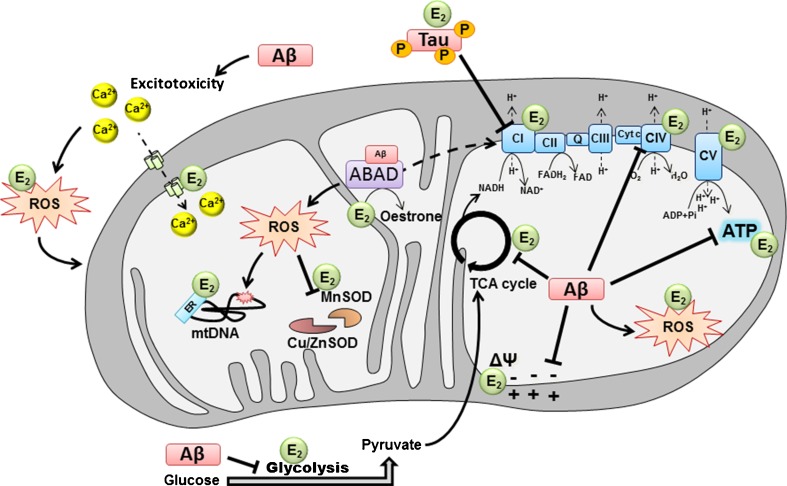
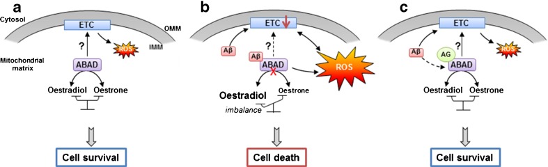
Hormonal deficit in post-menopausal women has been proposed to be one risk factor in Alzheimer's disease (AD) since two thirds of AD patients are women. However, large treatment trials showed negative effects of long-term treatment with oestrogens in older women. Thus, oestrogen treatment after menopause is still under debate, and several hypotheses trying to explain the failure in outcome are under discussion. Concurrently, it was shown that amyloid-beta (Aβ) peptide, the main constituent of senile plaques, as well as abnormally hyperphosphorylated tau protein, the main component of neurofibrillary tangles, can modulate the level of neurosteroids which notably represent neuroactive steroids synthetized within the nervous system, independently of peripheral endocrine glands. In this review, we summarize the role of neurosteroids especially that of oestrogen in AD and discuss their potentially neuroprotective effects with specific regard to the role of oestrogens on the maintenance and function of mitochondria, important organelles which are highly vulnerable to Aβ- and tau-induced toxicity. We also discuss the role of Aβ-binding alcohol dehydrogenase (ABAD), a mitochondrial enzyme able to bind Aβ peptide thereby modifying mitochondrial function as well as oestradiol levels suggesting possible modes of interaction between the three, and the potential therapeutic implication of inhibiting Aβ-ABAD interaction.


Conflict of interest statement

The authors declare that they have no conflict of interest.

Figures

Fig. 1
Fig. 1
Main biochemical pathways for neurosteroidogenesis in the vertebrate brain. Boxes represent neurosteroids which are sensitive to modulation by AD key proteins, Aβ and/or tau. Mitochondrial 17β-HSD (marked by *) is equivalent to the ABAD in mitochondria. PREG pregnenolone, PROG progesterone, 17OH-PREG 17-hydroxypregnenolone, 17OH-PROG 17-hydroxyprogesterone, DHEA dehydroepiandrosterone, DHP dihydroprogesterone, ALLOPREG allopregnanolone, DHT dihydrotestosterone, P450scc cytochrome P450 cholesterol side chain cleavage, P450c17 cytochrome P450c17, 3β-HSD 3β-hydroxysteroid dehydrogenase, 5α-R 5α-reductase, Arom. aromatase, 21-OHase 21-hydroxylase, 3α-HSOR 3α-hydroxysteroid oxydoreductase, 17β-HSD 17β-hydroxysteroid dehydrogenase
Fig. 2
Fig. 2
Modulation of mitochondrial function by Aβ, hyperphosphorylated tau and oestradiol. In AD, mitochondrial dysfunction was found to be a central pathological mechanism which occurs already at early stages of the disease. On one hand, studies showed that amyloid-β peptide (Aβ) can be responsible of metabolic impairments, such as the decrease of glucose consumption observed in the AD brain as well as the calcium-induced excitotoxicity in neurons. It has been found that hyperphosphorylated tau and Aβ are able to impair mitochondrial respiration by inhibiting the ETC CI and CIV, respectively, inducing decreased oxygen consumption, decreased ATP production and increased ROS level. This oxidative stress induced by ETC dysfunction can surpass cellular and mitochondrial scavenger (MnSOD, Cu/ZnSOD) and impacts on MMP as well as mitochondrial DNA (mtDNA). On the other hand, it has been shown that oestradiol can increase glucose utilization by cells as well as ETC activity, stabilize the MMP and prevent ROS production and calcium-induced excitotoxicity. In the graph, E 2 designates where oestradiol potentially acts on mitochondria to compensate Aβ-induced toxicity. In turn, Aβ seems to be able to impact oestradiol metabolism in mitochondria, since it can be directly linked to the mitochondrial enzyme ABAD and possibly modulates its enzymatic activity (such as the reversible conversion of oestradiol to oestrone) and non-enzymatic activity (mitochondrial RNAse P). ABAD Aβ-binding alcohol dehydrogenase, CI complex I, CII complex II, CIII complex III, CIV complex IV, CV complex V, cyt c cytochrome c, Cu/Zn SOD copper/zinc superoxide dismutase, MnSOD manganese superoxide dismutase, TCA tricyclic acid, E 2 oestradiol, ROS reactive oxygen species, mtDNA mitochondrial DNA, ER oestrogen receptor
Fig. 3
Fig. 3
Aβ, ABAD and mitochondria: modes of interactions. a Under normal conditions, ABAD is responsible of the reversible oxido/reduction of linear alcohols and steroids, such as the reversible conversion from oestradiol to oestrone. Its potential function as an RNAse P could also be important for the good functioning of the mitochondrial ETC. b Under AD-relevant pathological conditions, Aβ can directly bind the mitochondrial enzyme ABAD, changing the configuration of the enzyme which seems to inhibit its activity and creates an imbalance between oestradiol and oestrone. Aβ-induced ABAD misfolding can impact ETC functioning and increase, directly or indirectly, ROS production, which lead to cell death. c In the presence of AG18051 (AG), the binding of Aβ to ABAD is inhibited, normalizing oestradiol level, ROS production, ETC activity, and improves cell survival. ABAD Aβ-binding alcohol dehydrogenase, IMM inner mitochondrial membrane, OMM outer mitochondrial membrane

Similar articles

Cited by

References

    1. Corpechot C, Robel P, Axelson M, Sjovall J, Baulieu EE. Characterization and measurement of dehydroepiandrosterone sulfate in rat brain. Proc Natl Acad Sci USA. 1981;78(8):4704–4707. doi: 10.1073/pnas.78.8.4704. - DOI - PMC - PubMed
    1. Mensah-Nyagan AG, Do-Rego JL, Beaujean D, Luu-The V, Pelletier G, Vaudry H. Neurosteroids: expression of steroidogenic enzymes and regulation of steroid biosynthesis in the central nervous system. Pharmacol Rev. 1999;51(1):63–81. - PubMed
    1. Patte-Mensah C, Kibaly C, Boudard D, Schaeffer V, Begle A, Saredi S, Meyer L, Mensah-Nyagan AG. Neurogenic pain and steroid synthesis in the spinal cord. J Mol Neurosci. 2006;28(1):17–31. doi: 10.1385/JMN:28:1:17. - DOI - PubMed
    1. Plassart-Schiess E, Baulieu EE. Neurosteroids: recent findings. Brain Res Rev. 2001;37(1–3):133–140. doi: 10.1016/S0165-0173(01)00113-8. - DOI - PubMed
    1. Tsutsui K. Neurosteroids in the Purkinje cell: biosynthesis, mode of action and functional significance. Mol Neurobiol. 2008;37(2–3):116–125. doi: 10.1007/s12035-008-8024-1. - DOI - PubMed

Publication types