. 2012 Jul 3;53(8):4539-45.
doi: 10.1167/iovs.11-9300.

Patterns of ganglion cell complex and nerve fiber layer loss in nonarteritic ischemic optic neuropathy by Fourier-domain optical coherence tomography

Affiliations

Patterns of ganglion cell complex and nerve fiber layer loss in nonarteritic ischemic optic neuropathy by Fourier-domain optical coherence tomography

Divya Aggarwal et al. Invest Ophthalmol Vis Sci. .

Abstract

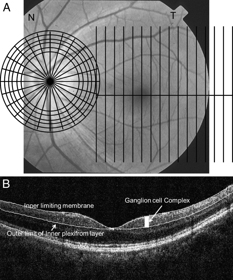
Purpose: To characterize by Fourier-domain optical coherence tomography (FD-OCT) the loss of nerve fiber layer (NFL) and ganglion cell complex (GCC) in nonarteritic ischemic optic neuropathy (NAION).

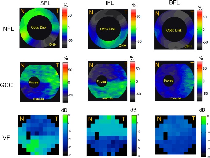
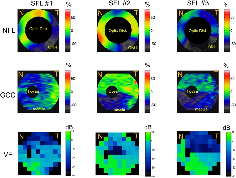
Methods: Patients diagnosed with NAION were enrolled and categorized into "superior field loss (SFL)," "inferior field loss (IFL)," and "bihemispheric field loss (BFL)" groups based on the Swedish interactive threshold algorithm 30-2 achromatic visual field (VF) tests. Six months after presentation, they were scanned by FD-OCT to map peripapillary NFL and macular GCC thicknesses. Age-matched normals were selected from participants in the Advanced Imaging for Glaucoma Study (www.AIGStudy.net). Deviation maps were defined as the difference between the thickness maps and the average normal maps. Pearson's correlation coefficient was used to assess the correlation between VF and OCT measurements.

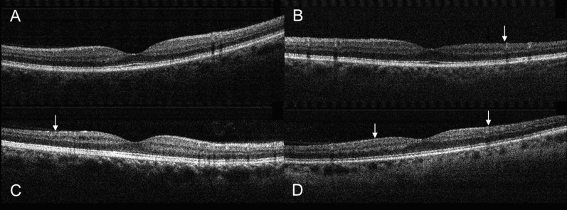
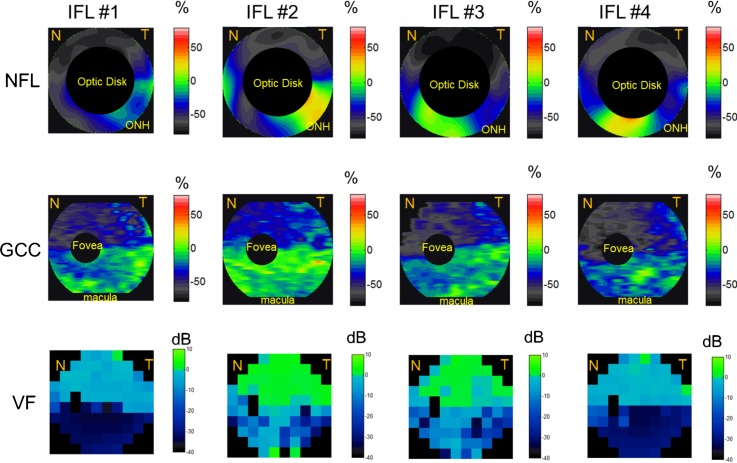
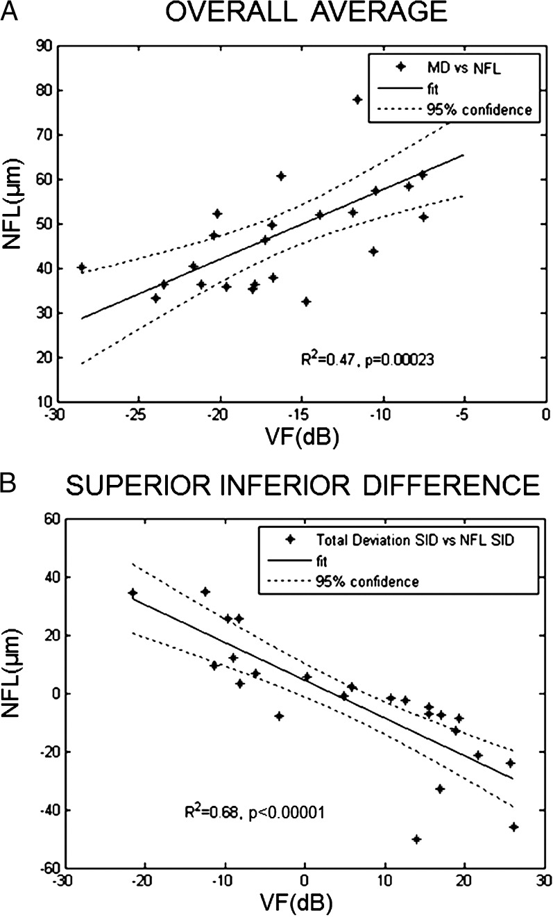
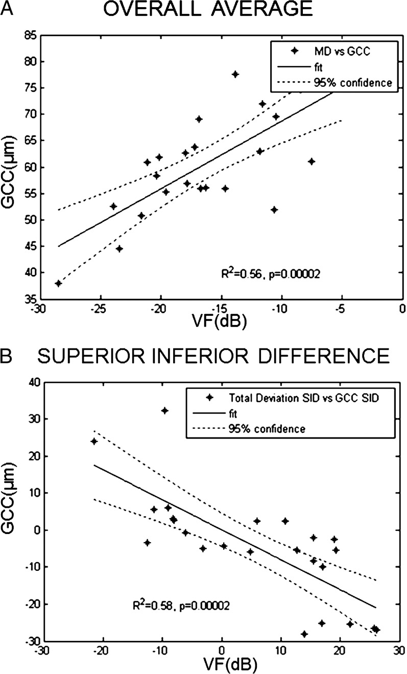
Results: Twenty-five NAION eyes in 20 subjects were analyzed. Most (2/3) SFL cases showed inferior NFL loss with variable sparing of inferonasal losses. All (4/4) IFL cases showed superior NFL loss with variable inferonasal extension. The GCC maps demonstrated clear hemispheric loss pattern in agreement with VFs. NFL and GCC losses could be detected even in the less affected hemispheres (P < 0.001). NFL and GCC were highly correlated (P < 0.001) with VF in terms of both overall averages and superior-inferior hemispheric differences.

Conclusions: NFL and GCC losses correlated well with VF losses in both magnitude and location. Hemispheric GCC loss correlated with altitudinal VF loss and this pattern may be of diagnostic value. FD-OCT is useful in the evaluation of NAION.


Conflict of interest statement

Disclosure: D. Aggarwal, None; O. Tan, Optovue (F, R), P; D. Huang, Optovue (F, C, R), P; A.A. Sadun, None

Figures

Figure 1
Figure 1
(A) The GCC and ONH scan patterns. The GCC scans consisted of 15 vertical line scans covering a 7 × 7 mm rectangular area temporal to fixation. The ONH scan patterns consisted of radial and circular scans on and around the ONH. The patterns were overlaid on a fundus photograph of a left eye. (B) OCT image overlaid with detected boundaries for GCC scan.
Figure 2
Figure 2
Vertical OCT scans centered on the fovea. (A) A normal eye. (B) An eye with SFL showed inferior thinning (arrow) of the GCC. (C) An eye with IFL showed superior GCC thinning (arrow). (D) An eye with BFL showed both superior and inferior GCC thinning (arrows). The examples shown were randomly picked from the three groups.
Figure 3
Figure 3
The average NFL, GCC, and VF loss patterns in the SFL, IFL, and BFL groups. SFL eyes demonstrated more loss of NFL and GCC in the inferior hemisphere and vice versa. Top row: NFL. Middle row: GCC. Bottom row: VF loss pattern. Left column: SFL group. Middle column: IFL group. Right column: BFL group. NFL loss map is around the 4-mm region of the optic disc, the GCC loss map covers the 7-mm macula region, and VF covers both ONH and macula nasal (N) and temporal (T) areas. Undefined regions of GCC loss, NFL loss, and VF were marked in black. Red and orange corresponded to GCC and NFL thickening; green corresponded to no loss; and blue and gray corresponded to GCC and NFL loss.
Figure 4
Figure 4
The NFL, GCC, and VF loss patterns of the three eyes with SFL. SFL eyes showed NFL and GCC loss predominantly in the inferior hemisphere. The NFL loss map is around the 4-mm region of the optic disc, the GCC loss map covers the 7-mm macula region, and VF covers both ONH and macula nasal (N) and temporal (T) areas. Undefined regions of GCC loss, NFL loss, and VF were marked in black. Red and orange corresponded to GCC and NFL thickening; green corresponded to no loss; and blue and gray corresponded to GCC and NFL loss.
Figure 5
Figure 5
The NFL, GCC, and VF loss patterns of all four eyes with IFL. IFL eyes showed NFL and GCC loss predominantly in the inferior hemisphere. The NFL loss map is around the 4-mm region of the optic disc, the GCC loss map covers the 7-mm macula region, and VF covers both ONH and macula nasal (N) and temporal (T) areas. Undefined regions of GCC loss, NFL loss, and VF were marked in black. Red and orange corresponded to GCC and NFL thickening; green corresponded to no loss; and blue and gray corresponded to GCC and NFL loss.
Figure 6
Figure 6
Correlation between NFL thickness and VF. (A) Mean deviation (MD) of VF test with average NFL thickness. (B) Superior–inferior difference (SID) of total deviation of VF test with SID of NFL thickness. For both MD and SID, there was a high degree of correlation between NFL thickness and VF.
Figure 7
Figure 7
Correlation between GCC thickness and VF. (A) MD of VF test with average GCC thickness. (B) SID of total deviation of VF test with SID of GCC thickness. For both MD and SID, there was a high degree of correlation between GCC thickness and VF.

Similar articles

Cited by

References

    1. Johnson LN, Arnold AC. Incidence of nonarteritic and arteritic anterior ischemic optic neuropathy. Population-based study in the state of Missouri and Los Angeles County, California. J Neuroophthalmol. 1994; 14: 38– 44. - PubMed
    1. Hattenhauer MG, Leavitt JA, Hodge DO, Grill R, Gray DT. Incidence of nonarteritic anterior ischemic optic neuropathy. Am J Ophthalmol. 1997; 123: 103– 107. - PubMed
    1. Levin LA, Louhab A. Apoptosis of retinal ganglion cells in anterior ischemic optic neuropathy. Arch Ophthalmol. 1996; 114: 488– 491. - PubMed
    1. Quigley HA, Miller NR, Green WR. The pattern of optic nerve fiber loss in anterior ischemic optic neuropathy. Am J Ophthalmol. 1985; 100: 769– 776. - PubMed
    1. Knox DL, Kerrison JB, Green WR. Histopathologic studies of ischemic optic neuropathy. Trans Am Ophthalmol Soc. 2000; 98: 202– 221. - PMC - PubMed

Publication types