. 2012 Nov;48(3):526-40.
doi: 10.1007/s12031-012-9818-1. Epub 2012 Jun 8.

Multimodal neuroprotection induced by PACAP38 in oxygen-glucose deprivation and middle cerebral artery occlusion stroke models

Affiliations

Multimodal neuroprotection induced by PACAP38 in oxygen-glucose deprivation and middle cerebral artery occlusion stroke models

Philip Lazarovici et al. J Mol Neurosci. 2012 Nov.

Abstract

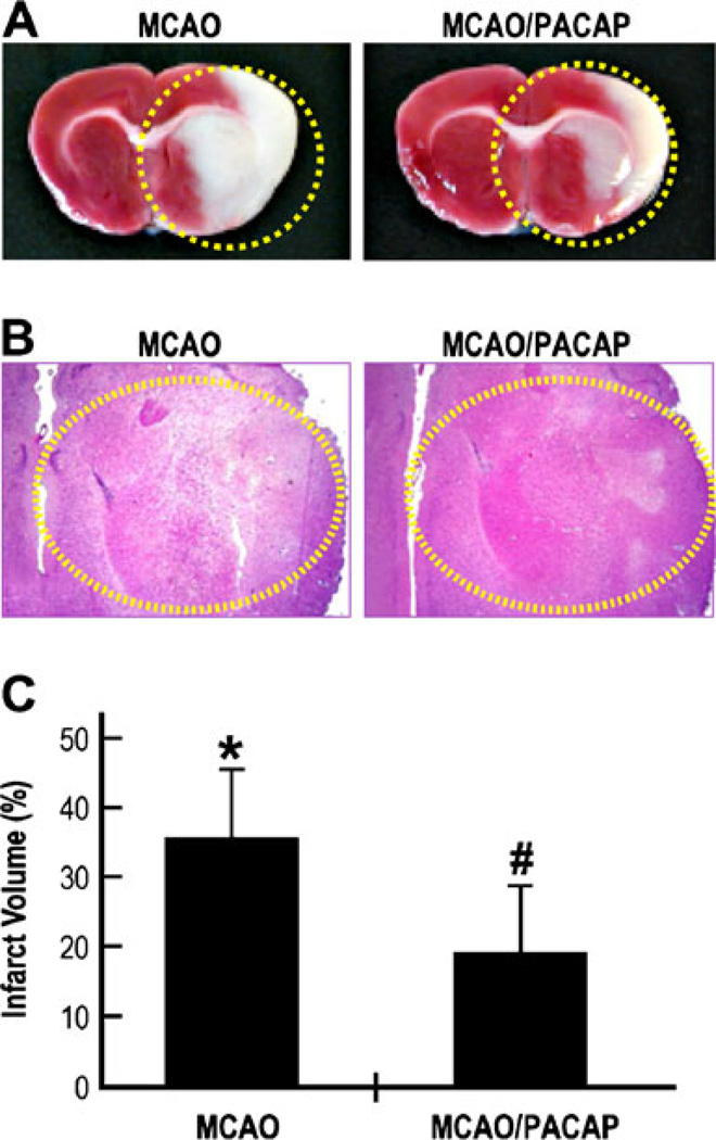
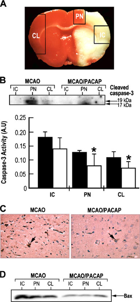
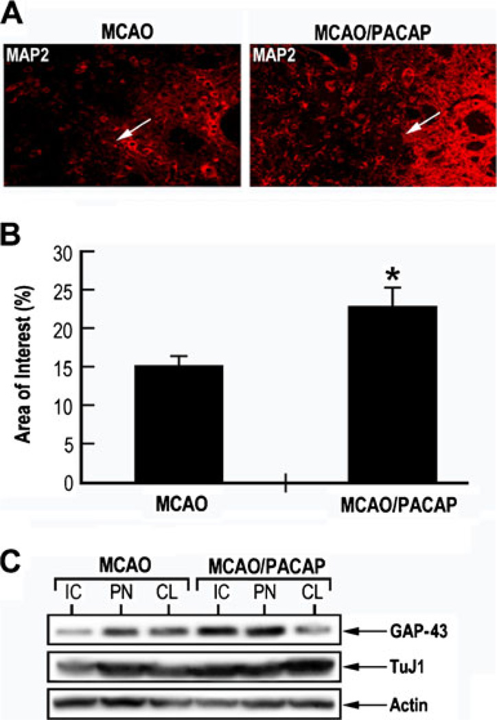
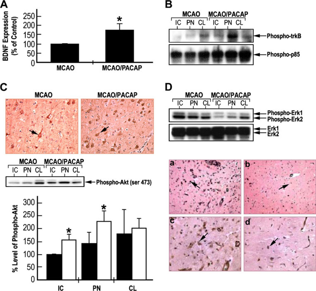
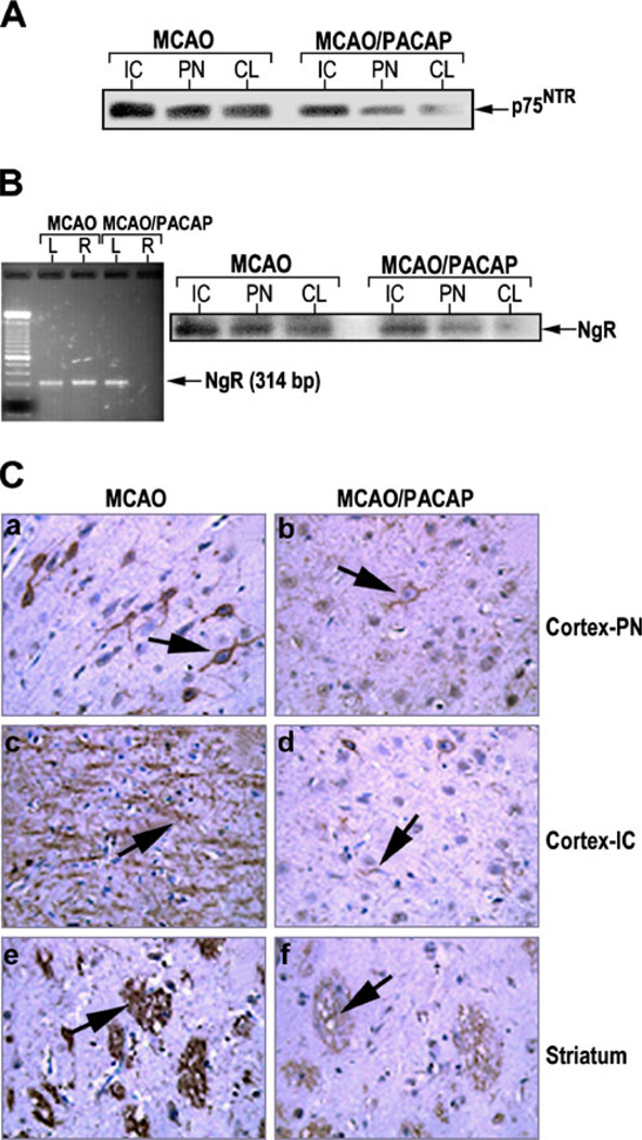
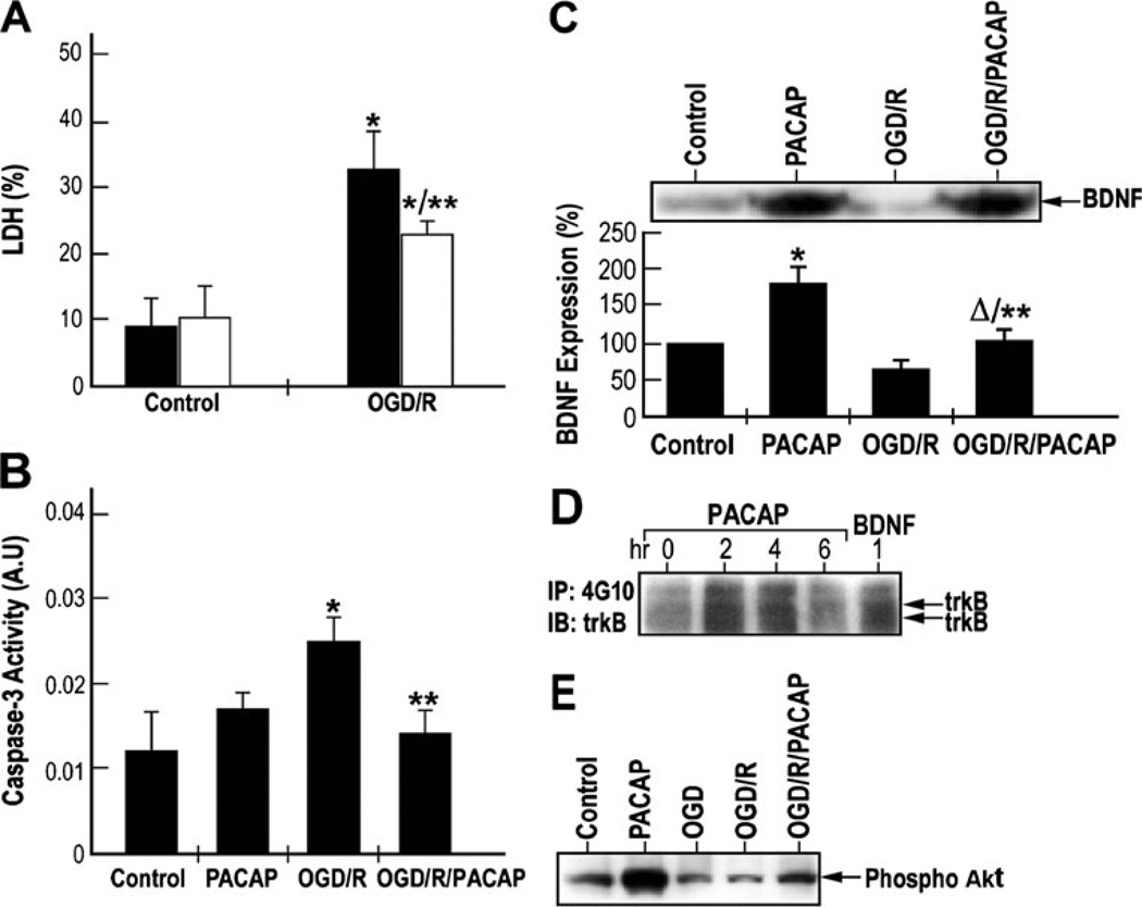
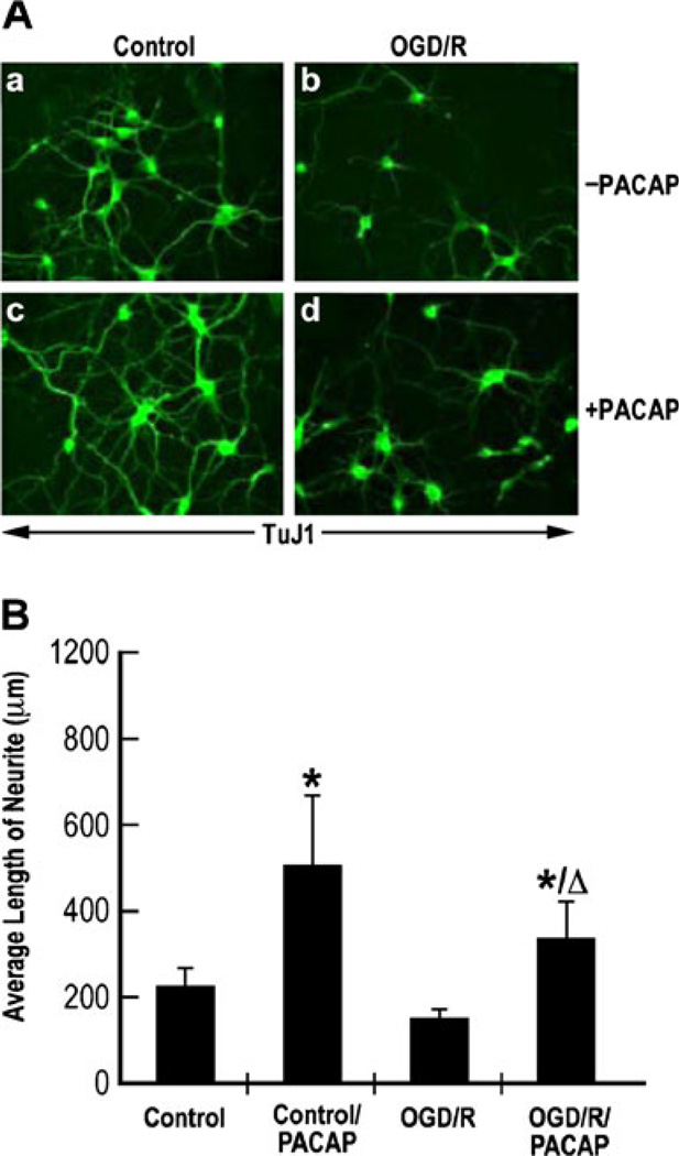
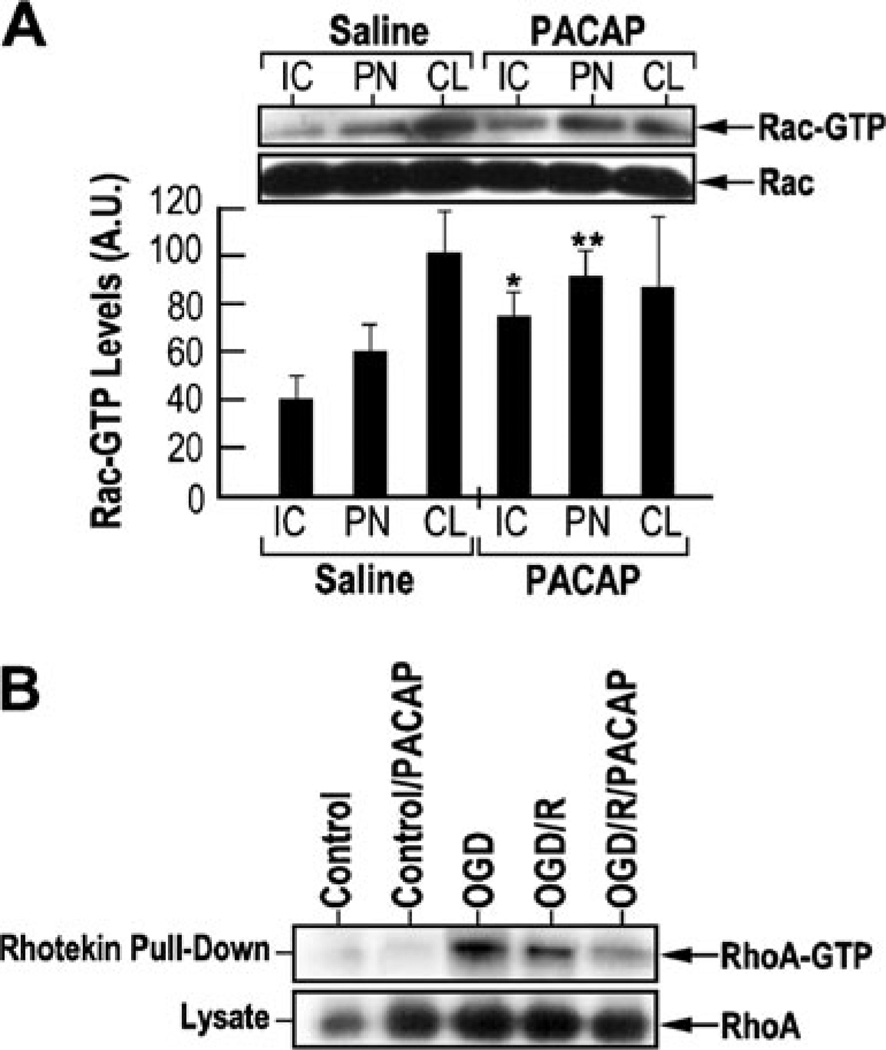
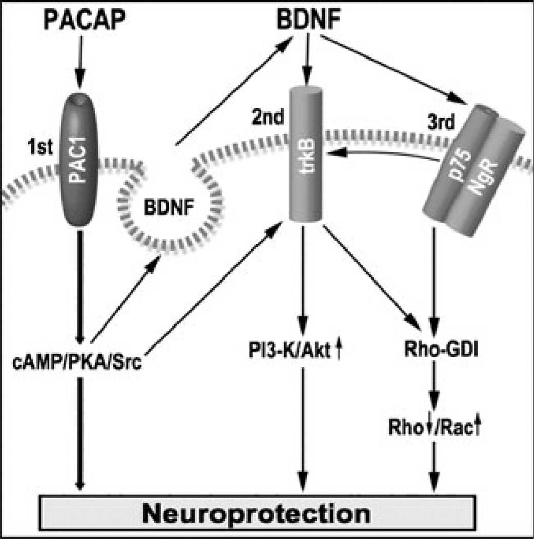
Pituitary adenylate cyclase activating peptide (PACAP), a potent neuropeptide which crosses the blood-brain barrier, is known to provide neuroprotection in rat stroke models of middle cerebral artery occlusion (MCAO) by mechanism(s) which deserve clarification. We confirmed that following i.v. injection of 30 ng/kg of PACAP38 in rats exposed to 2 h of MCAO focal cerebral ischemia and 48 h reoxygenation, 50 % neuroprotection was measured by reduced caspase-3 activity and volume of cerebral infarction. Similar neuroprotective effects were measured upon PACAP38 treatment of oxygen-glucose deprivation and reoxygenation of brain cortical neurons. The neuroprotection was temporally associated with increased expression of brain-derived neurotrophic factor, phosphorylation of its receptor-tropomyosin-related kinase receptor type B (trkB), activation of phosphoinositide 3-kinase and Akt, and reduction of extracellular signal-regulated kinases 1/2 phosphorylation. PACAP38 increased expression of neuronal markers beta-tubulin III, microtubule-associated protein-2, and growth-associated protein-43. PACAP38 induced stimulation of Rac and suppression of Rho GTPase activities. PACAP38 downregulated the nerve growth factor receptor (p75(NTR)) and associated Nogo-(Neurite outgrowth-A) receptor. Collectively, these in vitro and in vivo results propose that PACAP exhibits neuroprotective effects in cerebral ischemia by three mechanisms: a direct one, mediated by PACAP receptors, and two indirect, induced by neurotrophin release, activation of the trkB receptors and attenuation of neuronal growth inhibitory signaling molecules p75(NTR) and Nogo receptor.


Figures

Fig. 1
Fig. 1
Neuroprotective effects of PACAP after focal cerebral ischemia measured by reduced infarct volume. a Rats were subjected to 2 h of right MCAO followed by 48 h of reoxygenation in the presence or absence of PACAP38 injected i.v. at a dose of 30 ng/kg into the tail vein. The rats were allowed to survive for 2 days. Brains were dissected, perfused, fixed, and cut into sections of 2 mm. A series of adjacent 6-µm-thick sections were cut from the 2-mm block (bregma level +1.0 to +3.0) in the coronal plane and stained with a TTC and b hematoxylin and eosin; MCAO: saline-treated; MCAO/PACAP: PACAP38-treated. c The infarct volume represents the percentage of the area of lesion in the ipsilateral hemisphere vs. the corresponding area in the contralateral hemisphere. *p<0.01 compared to healthy brains; #p< 0.01 compared to MCAO brains
Fig. 2
Fig. 2
Neuroprotective effects of PACAP after focal cerebral ischemia measured by reduced caspase-3 and Bax (a member of the pro-apoptotic Bcl-2 protein family) expression. a Representative coronal brain sections (4 mm) stained with 2,3,5-triphenyltetrazolium chloride to demonstrate the regions of tissue dissection. Ipsilateral hemisphere: ischemic core (IC) and penumbra (PN) boundary zone to the ischemic core; contralateral hemisphere (CL). b PACAP reduces caspase-3 activity under ischemic conditions. Insert, western blotting analysis using the antibody against active caspase-3 and caspase-3 activity measured by a colorimetric kit. Black bars: saline; white bars: PACAP38; *p<0.05 compared to saline. c Immunostaining of coronal brain sections (6 µM) using anti-active caspase-3 antibody. d Western blotting analysis of Bax using the tissue samples from ischemic core (IC), penumbra (PN), and contralateral (CL) hemispheres. MCAO rats injected with either saline (MCAO) or PACAP38 (MCAO/PACAP)
Fig. 3
Fig. 3
PACAP induced MAP-2 TuJ1 and Gap-43 immunoreactivity in MCAO model. a Immunostaining of MAP-2 was performed on coronal brain sections from MCAO rats injected with either saline (MCAO) or PACAP38 (MCAO/PACAP), using a monoclonal antibody against MAP-2. b Quantitative analysis of MAP-2 immunopositive staining in the ischemic penumbra (PN) area. *p<0.05 compared to MCAO. c Western blotting analysis of different neuronal cytoskeletal markers. Brain tissue samples from MCAO rats injected with either saline (MCAO) or PACAP38 (MCAO/PACAP) were dissected and cell lysates were prepared. Equal amounts of lysates were immunoblotted using specific monoclonal antibodies against TuJ1, GAP-43, and actin. IC ischemic core, PN penumbra, CL contralateral
Fig. 4
Fig. 4
PACAP induced expression of BDNF and phosphorylation of trkB/PI3-K–Akt in MCAO rat model. a ELISA evaluation of BDNF expression in lysates from tissue samples from penumbra of ipsilateral hemisphere of the MCAO rats injected with either saline or PACAP38; *p<0.01 compared to MCAO. b PACAP-induced tyrosine phosphorylation of trkB and PI3-K p85. Equal amounts of tissue samples of the MCAO rats injected with either saline or PACAP38 were immunoprecipitated with anti-phosphotyrosine antibody and then immunoblotted with anti-trkB or anti-PI3-K p85 antibodies. c PACAP-induced Akt phosphorylation: upper part, immunostaining of coronal brain sections with anti-phospho-Akt antibody; middle part, western blotting analysis of phospho-Akt levels; lower part, the levels of phospho-Akt were measured from three western blotting and represented as mean±SD; black bars—saline; white bars—PACAP; *p<0.05 compared to saline in the same group. d PACAP reduced Erk1/2 phosphorylation. Upper part western blotting analysis was performed using antibodies against phospho-Erk1/2 and basal Erk1. Lower part immunostaining of the coronal brain sections using anti-phospho-Erk1/2. a, b Low magnification (×20); c, d high magnification (×40); a, c—MCAO; b, d— MCAO/PACAP. IC ischemic core, PN penumbra, CL contralateral
Fig. 5
Fig. 5
PACAP reduction of p75NTR/NgR expression in MCAO rat model. a PACAP mediated downregulation of p75NTR and b down-regulation of Nogo receptor (NgR). The tissues were dissected from ischemic core (IC) and penumbra (PN) areas of the ipsilateral hemisphere, as well as the corresponding region of the contralateral (CL) hemisphere, from the rats subjected to MCAO or MCAO followed by PACAP38 treatment. Equal amounts of lysates were subjected to western blotting analysis using anti-p75NTR or anti-NgR receptor antibodies. b Left—PACAP-mediated downregulation of Nogo receptors measured by RT-PCR. L: tissue sample from contralateral hemisphere; R: tissue sample from ipsilateral hemisphere. Total RNA was isolated and RT-PCR was performed at 40 cycles at 94 °C for 1 min, 62 °C for 1 min, and 72 °C for 1 min. Samples were separated on 2 % agarose gel. c Immunostaining of coronal brain sections with anti-Nogo receptor antibody indicating PACAP downregulation of Nogo receptor expression. The 6-µm-thick sections were derived from the 2-mm brain block D (bregma level +1.0 to +3.0) stained with anti-Nogo receptor antibody. a, b Cortex at penumbra; c, d cortex at ischemic core; e, f striatum. Arrows indicate NgR positive cells
Fig. 6
Fig. 6
PACAP induced neuroprotection, BDNF release, and trkB and Akt phosphorylation in cultured cortical neurons subjected to oxygen–glucose deprivation (OGD). a Neuroprotective effects of PACAP on OGD-induced neuronal cell death in vitro. Cultured cortical neurons were subjected to OGD for 1 h followed by reperfusion for 48 h, in the absence (OGD/R) or presence (OGD/R/PACAP) of 100 nM of PACAP38. The levels of released and total lactate dehydrogenase (LDH) were measured by using a CytoTox 96 cytotoxicity assay kit. Results were presented as mean±SD, *p<0.05 compared to control; **p<0.05 compared to the saline group exposed to oxygen–glucose deprivation followed by reoxygenation (OGD/R) (black—saline; white —PACAP38). b Inhibitory effects of PACAP on caspase-3 activity under OGD followed by reperfusion. Cultured cortical neurons were subjected to OGD for 1 h followed by reperfusion as in a. Caspase-3 activity was measured using caspase-3 assay kit. Results were presented as mean±SD. *p <0.05 compared to control; **p <0.05 compared to OGD/R. c Upper—western blotting analysis of BDNF expression. Lower—quantization of BDNF levels using ELISA. *p< 0.001 compared to control; **p<0.001 compared to OGD/R; Δp<0.05 compared to PACAP. d Effects of PACAP on the activation of trkB-mediated signaling pathways in cultured cortical neurons. Cultured cortical neurons were treated with 100 nM of PACAP38 for 2, 4, and 6 h. Cells were also treated with 50 ng/ml of BDNF for 1 h. Equal amounts of lysates were immunoprecipitated with anti-phosphotyrosine antibody and immunoblotted with anti-trkB or anti-PI3-K p85 antibodies. e PACAP induces Akt phosphorylation under ischemic conditions. Cultured cortical neurons were subjected to OGD for 1 h followed by reperfusion for 48 h, in the absence or presence of 100 nM of PACAP38. Equal amounts of lysates were subjected to western blotting analysis. Phosphorylation of Akt was detected by using an anti-phospho-Akt antibody (ser 43)
Fig. 7
Fig. 7
PACAP induced neurite outgrowth under ischemic conditions in cultured cortical neurons subjected to OGD. a Immunostaining of cultured cortical neurons with TuJ1. Cultured cortical neurons subjected to OGD followed by reoxygenation (OGD/R) or exposed to normoxic conditions (control) were left untreated (−PACAP; a, b) or treated with 100 nM of PACAP38 for 48 h (+PACAP; c, d). Cells were fixed and subjected to immunostaining using TuJ1 antibody. b Quantization of the average length of neurite from different conditions shown in a. Photomicrographs were taken at × 40 magnification with a digital camera. The axons were then traced and measured using SigmaScan analysis system. Results were presented as mean±SD. *p<0.05 as compared to control; Δp<0.05 as compared to oxygen–glucose deprivation followed by reoxygenation (OGD/R)
Fig. 8
Fig. 8
PACAP induction of Rac1 (Ras-related C3 botulinum toxin substrate 1) and suppression of RhoA (Ras homolog gene family, member A) activity. a The effect of PACAP on Rac activity. Insert— equal amounts of lysates from the brain areas of rats subjected to MCAO or MCAO followed by PACAP38 treatment were incubated with PAK-1-PBD (p21-binding domain of human PAK-1). Precipitated Rac-GTP proteins were separated on 14 % SDS polyacrylamide gel and immunoblotted with anti-Rac1 antibody. Equal amounts of lysates were run separately to measure the total Rac levels. Quantization of Rac-GTP levels in various brain regions with or without PACAP treatment, using the data obtained from western blotting experiments. IC ischemic core, PN penumbra, CL contralateral. b The effect of PACAP RhoA activity in cultured cortical neurons subjected to 1 h of OGD or 1 h of OGD followed by 1 h of reperfusion, in the absence or presence of 100 nM PACAP38. RhoA-GTP activity was measured using a Rhotekin pull-down assay kit. Equal amounts of lysates were subjected to immunoblotting to measure the total RhoA levels among different samples. Control—normoxia; control/PACAP—normoxia and 100 nM PACAP treatment; oxygen–glucose deprivation—OGD alone; OGD/R—OGD followed by 48 h of reoxygenation; OGD/R/PACAP—OGD/R with 100 nM PACAP treatment
Fig. 9
Fig. 9
Schematic diagram of potential crosstalk signaling pathway between PACAP and BDNF leading to neuroprotection; 1st, 2nd, and 3rd represent the three mechanisms leading to neuroprotection; arrow up—activation; arrow down—inhibition; striped line—plasma membrane

Similar articles

Cited by

References

    1. Arien-Zakay H, Lecht S, Bercu MM, et al. Neuroprotection by cord blood neural progenitors involves antioxidants, neurotrophic and angiogenic factors. Exp Neurol. 2009;216:83–94. - PubMed
    1. Arimura A. Perspectives on pituitary adenylate cyclase activating polypeptide (PACAP) in the neuroendocrine, endocrine, and nervous systems. Jpn J Physiol. 1998;48:301–331. - PubMed
    1. Benowitz LI, Perrone-Bizzozero NI, Neve RL, Rodriguez W. GAP-43 as a marker for structural plasticity in the mature CNS. Prog Brain Res. 1990;86:309–320. - PubMed
    1. Bito H, Takemoto-Kimura S. Ca(2+)/CREB/CBP-dependent gene regulation: a shared mechanism critical in long-term synaptic plasticity and neuronal survival. Cell Calcium. 2003;34:425–430. - PubMed
    1. Botia B, Basille M, Allais A, et al. Neurotrophic effects of PACAP in the cerebellar cortex. Peptides. 2007;28:1746–1752. - PubMed

Publication types

MeSH terms

Substances