. 2012 Aug 1;303(3):G344-55.
doi: 10.1152/ajpgi.00403.2011. Epub 2012 Jun 7.

Cytosolic sulfotransferase 2B1b promotes hepatocyte proliferation gene expression in vivo and in vitro

Affiliations

Cytosolic sulfotransferase 2B1b promotes hepatocyte proliferation gene expression in vivo and in vitro

Xin Zhang et al. Am J Physiol Gastrointest Liver Physiol. .

Abstract

Cytosolic sulfotransferase 2B1b (SULT2B1b) catalyzes the sulfation of 3β-hydroxysteroids and functions as a selective cholesterol and oxysterol sulfotransferase. Activation of liver X receptors (LXRs) by oxysterols has been known to be an antiproliferative factor. Overexpression of SULT2B1b impairs LXR's response to oxysterols, by which it regulates lipid metabolism. The aim of this study was to investigate in vivo and in vitro effects of SULT2B1b on liver proliferation and the underlying mechanisms. Primary rat hepatocytes and C57BL/6 mice were infected with adenovirus encoding SULT2B1b. Liver proliferation was determined by measuring the proliferating cell nuclear antigen (PCNA) immunostaining labeling index. The correlation between SULT2B1b and PCNA expression in mouse liver tissues was determined by double immunofluorescence. Gene expressions were evaluated by quantitative real-time PCR and Western blot analysis. SULT2B1b overexpression in mouse liver tissues increased PCNA-positive cells in a dose- and time-dependent manner. The increased expression of PCNA in mouse liver tissues was only observed in the SULT2B1b transgenic cells. Small interference RNA SULT2B1b significantly inhibited cell cycle regulatory gene expressions in primary rat hepatocytes. LXR activation by T0901317 effectively suppressed SULT2B1b-induced gene expression in vivo and in vitro. SULT2B1b may promote hepatocyte proliferation by inactivating oxysterol/LXR signaling.


Figures

Fig. 1.
Fig. 1.
Sulfotransferase 2B1b (SULT2B1b) mRNA and protein levels in mouse liver tissues following infection. Mice were infected with Ad-Control (Con) or Ad-SULT2B1b (Sult). On days 0, 2, 4, 5, and 12, mice were euthanized and liver tissues were collected. A: relative mRNA level of SULT2B1b expression measured by RT-PCR. An equivalent of total RNA for each sample was loaded, and 25 cycles for each sample were used for PCR analysis. B: Western blot analysis of SULT2B1b protein level.
Fig. 2.
Fig. 2.
Effect of SULT2B1b on proliferating cell nuclear antigen (PCNA) expression in mouse liver tissues. Mice were infected with Ad-Control (Con) or Ad-SULT2B1b (Sult). On days 0, 2, 4, 5, and 12, mice were euthanized and liver tissues were harvested. A: representative photomicrographs from PCNA-stained liver sections (×20 optical field). B: percentage of PCNA-positive cells obtained from liver sections. Values are means ± SD (n = 3–5/group) *P < 0.05 vs. day 0. C: Western blot analysis of SULT2B1b and PCNA protein levels.
Fig. 3.
Fig. 3.
Immunofluorescent double-labeled images illustrate locations of SULT2B1b (red) and PCNA (green) proteins in mouse liver tissues. A–C: in a liver section of mouse infected with Ad-SULT2B1b for 4 days, blue fluorescence shows all cell nuclei with patterns of 4′,6-diamidino-2-phenylindole (DAPI) staining (A), red fluorescence shows cytoplasmic expression of SULT2B1b (B), and green fluorescence shows nuclear expression of PCNA (C). D: merged image shows that hepatocytes with PCNA expression in the nuclei are accompanied by SULT2B1b overexpression in the cytosol, while there was no PCNA expression in cells without SULT2B1b overexpression. Images represent results from 3 separate experiments.
Fig. 4.
Fig. 4.
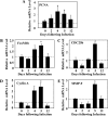
Effect of SULT2B1b overexpression on proliferative gene expressions in mouse liver tissues. After infection with Ad-Control or Ad-SULT2B1b, mice were euthanized on days 0, 2, 4, 5, and 12. Total mRNAs were prepared from liver tissues, and each mRNA level was analyzed by real-time quantitative PCR. A–E: PCNA, forkhead box M1b (FoxM1b), cell division cycle 25b (CDC25b), cyclin A, and matrix metalloproteinase 9 (MMP-9) expressions at mRNA level. Values are means ± SD (n = 3–5/group). *P < 0.05 vs. day 0.
Fig. 5.
Fig. 5.
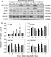
Effect of SULT2B1b overexpression on liver X receptor (LXR) and its target gene expressions in mouse liver tissues. Mice were euthanized on days 0, 2, 4, 5, and 12 following Ad-Control or Ad-SULT2B1b infection. A and B: Western blot analysis of hepatic SULT2B1b, PCNA, LXRα, ATP-binding cassette transporter 1 (ABCA1), and sterol regulatory element-binding protein (SREBP)-1 protein levels in control (black bars) and SULT2B1b (gray bars) groups. Values are means ± SDqj (n = 3–5/group). #P < 0.05 vs. Con.
Fig. 6.
Fig. 6.
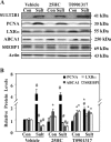
Role of LXR signaling in SULT2B1b-induced proliferation in mouse liver tissues. After infection with Ad-Control or Ad-SULT2B1b for 24 h, mice were treated with 25-hydroxycholesterol (25HC) or T0901317 (25 mg/kg ip) for 24 h and euthanized on day 5. A and B: Western blot analysis of hepatic protein levels of SULT2B1b, PCNA, LXR, and its target genes ABCA1 and SREBP1. Values are means ± SD (n = 3–5/group). *P < 0.05 vs. corresponding vehicle. #P < 0.05 vs. Con.
Fig. 7.
Fig. 7.
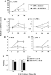
Effect of small interference RNA (siRNA)-SULT2B1b on proliferation in primary rat hepatocytes (PRH). PRH were transfected with siRNA-SULT2B1b or siRNA-Control for 24 and 48 h. A: real-time quantitative PCR analysis of SULT2B1b mRNA level after siRNA-SULT2B1b transfection. B–E: real-time quantitative PCR analysis of cell cycle regulatory gene [PCNA, FoxM1b, cyclin-dependent kinase 2 (CDK2), and cyclin A] mRNA levels. F: cell survival assay of relative hepatocyte viability. Optical density value of cells cultured in normal medium (N) was arbitrarily set at 100%. Values are means ± SD of 3 determinations. *P < 0.05 vs. N. #P < 0.05 vs. siRNA-Control.
Fig. 8.
Fig. 8.
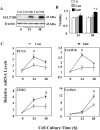
Effect of SULT2B1b overexpression on proliferation in PRH. PRH were infected with Ad-Control or Ad-SULT2B1b for 24 and 48 h. A: Western blot analysis of SULT2B1b protein level. B: cell survival assay of relative hepatocyte viabilities. Optical density value of cells cultured in normal medium (N) was arbitrarily set at 100%. C: real-time quantitative PCR analysis of cell cycle regulatory gene (PCNA, FoxM1b, CDK2, and cyclin A) mRNA levels. Values are means ± SD of 3 determinations. *P < 0.05 vs. N. #P < 0.05 vs. Con.
Fig. 9.
Fig. 9.
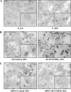
Effect of SULT2B1b on PCNA expression in PRH. PRH were plated on glass coverslips and infected with adenovirus or siRNA. A–C: PCNA-positive cells in hepatocytes cultured in normal medium (N_0 h and N_48 h) or transfected with adenovirus (Ad-Control_48 h and Ad-SULT2B1b_48 h) or siRNA (siRNA-Control_48 h and siRNA-SULT2B1b_48 h) for 48 h were examined by immunocytochemistry. Images represent randomly selected images from 3 different dishes, repeated in 3 independent experiments. Original magnification: ×20 and ×40 (insets).
Fig. 10.
Fig. 10.
Effect of SULT2B1b on proliferation via LXR signaling pathway in PRH. PRH were infected with Ad-Control or Ad-SULT2B1b for 24 and 48 h. A and B: Western blot analysis of SULT2B1b, PCNA, LXRα, ABCA1, and SREBP1 protein levels. C and D: at 24 h after infection, cells were treated with 25HC (3 μM) or T0901317 (1.5 μM) for 24 h, and protein levels of SULT2B1b, PCNA, and LXR and its target genes ABCA1 and SREBP1 were analyzed by Western blot. Blots represent results from 1 of 3 separate experiments. Values are means ± SD of 3 determinations. *P < 0.05 vs. 0 h. #P < 0.05 vs. Con.

References

    1. Bai Q, Xu L, Kakiyama G, Runge-Morris MA, Hylemon PB, Yin L, Pandak WM, Ren S. Sulfation of 25-hydroxycholesterol by SULT2B1b decreases cellular lipids via the LXR/SREBP-1c signaling pathway in human aortic endothelial cells. Atherosclerosis 214: 350– 356, 2011 - PMC - PubMed
    1. Bensinger SJ, Bradley MN, Joseph SB, Zelcer N, Janssen EM, Hausner MA, Shih R, Parks JS, Edwards PA, Jamieson BD, Tontonoz P. LXR signaling couples sterol metabolism to proliferation in the acquired immune response. Cell 134: 97– 111, 2008 - PMC - PubMed
    1. Bjorkhem I. Do oxysterols control cholesterol homeostasis? J Clin Invest 110: 725– 730, 2002 - PMC - PubMed
    1. Blaschke F, Leppanen O, Takata Y, Caglayan E, Liu J, Fishbein MC, Kappert K, Nakayama KI, Collins AR, Fleck E, Hsueh WA, Law RE, Bruemmer D. Liver X receptor agonists suppress vascular smooth muscle cell proliferation and inhibit neointima formation in balloon-injured rat carotid arteries. Circ Res 95: e110– e123, 2004 - PubMed
    1. Bochelen D, Mersel M, Behr P, Lutz P, Kupferberg A. Effect of oxysterol treatment on cholesterol biosynthesis and reactive astrocyte proliferation in injured rat brain cortex. J Neurochem 65: 2194– 2200, 1995 - PubMed

Publication types

LinkOut - more resources