. 2012 Aug 1;186(3):261-72.
doi: 10.1164/rccm.201201-0164OC. Epub 2012 Jun 7.

Modern age pathology of pulmonary arterial hypertension

Affiliations

Modern age pathology of pulmonary arterial hypertension

Elvira Stacher et al. Am J Respir Crit Care Med. .

Abstract

Rationale: The impact of modern treatments of pulmonary arterial hypertension (PAH) on pulmonary vascular pathology remains unknown.

Objectives: To assess the spectrum of pulmonary vascular remodeling in the modern era of PAH medication.

Methods: Assessment of pulmonary vascular remodeling and inflammation in 62 PAH and 28 control explanted lungs systematically sampled.

Measurements and main results: Intima and intima plus media fractional thicknesses of pulmonary arteries were increased in the PAH group versus the control lungs and correlated with pulmonary hemodynamic measurements. Despite a high variability of morphological measurements within a given PAH lung and among all PAH lungs, distinct pathological subphenotypes were detected in cohorts of PAH lungs. These included a subset of lungs lacking intima or, most prominently, media remodeling, which had similar numbers of profiles of plexiform lesions as those in lungs with more pronounced remodeling. Marked perivascular inflammation was present in a high number of PAH lungs and correlated with intima plus media remodeling. The number of profiles of plexiform lesions was significantly lower in lungs of male patients and those never treated with prostacyclin or its analogs.

Conclusions: Our results indicate that multiple features of pulmonary vascular remodeling are present in patients treated with modern PAH therapies. Perivascular inflammation may have an important role in the processes of vascular remodeling, all of which may ultimately lead to increased pulmonary artery pressure. Moreover, our study provides a framework to interpret and design translational studies in PAH.


Figures

Figure 1.
Figure 1.
Demographic characteristics of the study population (control subjects, n = 28; patients with pulmonary arterial hypertension [PAH], n = 62). (A) Distribution of sex in relation to age (displayed in decades) in control subjects and patients with PAH (solid, men; shaded, women). (B) Distribution of mean pulmonary arterial pressure (mPAP) in PAH (n = 62). (C) Distribution of pulmonary vascular resistance (PVR) in PAH (n = 60).
Figure 2.
Figure 2.
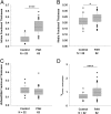
Assessment of intima fractional thickness (A), media fractional thickness (B), and adventitia fractional thickness (C) in lungs of control subjects (n = 22) and patients with pulmonary arterial hypertension (PAH) (n = 62). Measurements were made using digital imaging analysis of circular profiles of pulmonary arteries. Control cases with vascular remodeling (n = 6) were excluded from this analysis and are shown in Figure E3. (Student t test, *P < 0.05, ****P < 0.001). (D) Volume density vessel thickness (intima plus media) weighed by airway septa (Vintima+media)/septa (Wilcoxon rank-sum test, P < 0.001).
Figure 3.
Figure 3.
Frequency distribution of intima (A), media (B), and adventitia (C) fractional thickness among control (n = 22, solid bars) and pulmonary arterial hypertension (PAH) lungs (n = 62, shaded bars) (A, Wilcoxon rank-sum test, P < 0.001; B, Student t test, P = 0.026; C, Student t test, P = 0.418).
Figure 4.
Figure 4.
Correlation of intima plus media fractional thickness in patients with pulmonary arterial hypertension (PAH) with (A) mean pulmonary arterial pressure (mPAP) (n = 62, P = 0.066, R = 0.235), and (B) pulmonary vascular resistance (PVR) (n = 60, P = 0.057, R = 0.247). Correlation of media fractional thickness in patients with PAH with (C) mPAP (n = 62, P = 0.036, R = 0.267), and (D) PVR (n = 60, P = 0.047, R = 0.258).
Figure 5.
Figure 5.
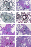
Characteristic histopathological findings encountered in idiopathic pulmonary arterial hypertension–like pattern. (A) Intima (arrows) and media (arrowheads) thickening (Russel-Movat pentachrome stain). (B) Cushion-like eccentric intima thickening (arrow) in pulmonary arteries (hematoxylin and eosin [H&E]). (C) Concentric “onion-skin–like” (arrow) intima thickening with subtotal luminal occlusion (Russel-Movat pentachrome stain). (D) Muscularization of small artery (arrow) (Russel-Movat pentachrome stain). (E) Plexiform lesion (arrow) fed by muscular artery (arrowheads) (H&E). (F) Subpleural plexiform lesion (arrow) (H&E). (G) Angiomatoid lesion consisting of dilated vessels (arrows) (H&E). (H) Recent thrombus (arrow) (H&E). Scale bars = 100 μm.
Figure 6.
Figure 6.
Characteristic histopathological findings observed in associated pulmonary arterial hypertension–collagen vascular disease–like pattern. (A) Fibrosis (arrow) and pronounced interstitial inflammation (arrowhead) (Russel-Movat pentachrome stain). (B) Media thickening of numerous arteries (arrows) and scant interstitial inflammation (hematoxylin and eosin [H&E]). (C) Fibroblastic focus (arrow), fibrosis (arrowheads), scant interstitial inflammation, and pronounced media thickening of an artery (H&E). (D) Architectural distortion with enlarged cyst-like lung spaces lined by respiratory epithelium (honeycombing) and focal moderate interstitial inflammation (arrowhead) (H&E). (E) Muscularized small pulmonary arteries (arrow) (H&E). (F) Peripheral plexiform lesion (arrow) within angiomatoid lesion (arrowhead) (H&E). Scale bars = 100 μm.
Figure 7.
Figure 7.
Comparison of hemodynamics with histologic pattern: (A) mean pulmonary arterial pressure (mPAP) (analysis of variance [ANOVA], P = not significant [NS]), and (B) pulmonary vascular resistance (PVR) (ANOVA, P = 0.008). Comparison of hemodynamics within the idiopathic pulmonary arterial hypertension (IPAH)-like group according to the underlying cause: (C) mPAP (ANOVA, P = NS), and (D) PVR (ANOVA, P = NS). **P < 0.01. APAH = associated PAH; CHD = congenital heart disease; FPAH = familial PAH; VOD = venoocclusive disease.
Figure 8.
Figure 8.
Measurements of (A) intima fractional thickness (analysis of variance [ANOVA], P < 0.001), (B) media fractional thickness (ANOVA, P = NS), and (C) adventitia fractional thickness (ANOVA, P = NS) in control subjects (CTL) and in patients with pulmonary arterial hypertension (PAH) on stratification into different histologic subgroups (idiopathic PAH [IPAH]-like pattern, associated PAH [APAH]–collagen vascular disease [CVD]-like pattern, or venoocclusive disease [VOD]-like pattern). Measurements were made using digital imaging of circular profiles of pulmonary arteries. (D) Measurement of intima fractional thickness in familial PAH and IPAH lungs of patients with (n = 4) or without identified BMPRII mutations (n = 34; Student t test). *P < 0.05, ****P < 0.001.
Figure 9.
Figure 9.
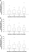
Comparison between plexiform lesions and vascular wall remodeling. Average number of profiles of plexiform lesions in correlation with quartiles of (A) intima fractional thickness, (B) media fractional thickness, and (C) adventitia fractional thickness in idiopathic PAH (IPAH)-like pattern and associated PAH (APAH)–collagen vascular disease (CVD)-like pattern (n = 60, n = 15/quartile).
Figure 10.
Figure 10.
(A) Coefficient of variation (CV), calculated as the standard deviation divided by the mean, as a measurement of variability of intima fractional thickness, media fractional thickness, and adventitia fractional thickness per slide in diseased patients (n = 62). (B) CV of profiles of plexiform lesions per slide in patients with plexiform lesions (n = 56). (C) Distribution of profiles of plexiform lesions per slide in idiopathic PAH (IPAH)-like pattern, each column on the x axis representing one patient (n = 48). Each column projecting from the x axis represents one individual patient; the respective y-dots represents the count of plexiform lesion profiles (i.e., outlines) in each of the 12 slides examined in each patient (P = not significant for all).
Figure 11.
Figure 11.
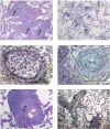
Examples of inflammation in pulmonary arterial hypertension (PAH) lungs. (A) Diffuse pronounced interstitial inflammation (arrows) (hematoxylin and eosin [H&E]). (B) Localized moderate perivascular inflammation (arrowheads) (H&E). (C) Occluded artery with dense inflammatory infiltrate (arrow) (Russel-Movat pentachrome stain). (D) Artery with concentric intima proliferation, neighbored by a dense inflammatory infiltrate (arrow) (Russel-Movat pentachrome stain). (E) Artery with plexiform lesion (arrow), embedded in inflammatory infiltrate (H&E). (F) Arteries focally cuffed by dense inflammation (arrows) (Russel-Movat pentachrome stain). Scale bars = 100 μm.
Figure 12.
Figure 12.
Correlation of the average perivascular inflammation score with (A) intima plus media fractional thickness (P = 0.042, R = 0.259), (B) adventitia fractional thickness (P = 0.030, R = 0.275), and (C) mean pulmonary arterial pressure (mPAP) (P = 0.075, R = 0.228) in pulmonary arterial hypertension (n = 62). The measurement of the average perivascular inflammation score is described in the online supplement.
Figure 13.
Figure 13.
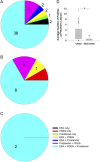
Use of pulmonary arterial hypertension (PAH)-specific medication in (A) idiopathic PAH–like pattern, (B) associated PAH–collagen vascular disease–like pattern, and (C) venoocclusive disease–like pattern. (D) Average number of profiles of plexiform lesions in PAH (n = 62) in correlation with use of prostanoids (Wilcoxon rank-sum test, P = 0.010). ERA = endothelin receptor antagonist. PDE5i = phosphodiesterase type 5 inhibitor.

Comment in

Similar articles

Cited by

References

    1. Humbert M, Sitbon O, Simonneau G. Treatment of pulmonary arterial hypertension. N Engl J Med 2004;351:1425–1436 - PubMed
    1. D’Alonzo GE, Barst RJ, Ayres SM, Bergofsky EH, Brundage BH, Detre KM, Fishman AP, Goldring RM, Groves BM, Kernis JT, et al. Survival in patients with primary pulmonary hypertension. Results from a national prospective registry. Ann Intern Med 1991;115:343–349 - PubMed
    1. Heath D, Edwards JE. The pathology of hypertensive pulmonary vascular disease; a description of six grades of structural changes in the pulmonary arteries with special reference to congenital cardiac septal defects. Circulation 1958;18:533–547 - PubMed
    1. Wagenvoort CA, Wagenvoort N. Primary pulmonary hypertension: a pathologic study of the lung vessels in 156 clinically diagnosed cases. Circulation 1970;42:1163–1184
    1. Rabinovitch M, Haworth SG, Castaneda AR, Nadas AS, Reid LM. Lung biopsy in congenital heart disease: a morphometric approach to pulmonary vascular disease. Circulation 1978;58:1107–1122 - PubMed

Publication types

MeSH terms