. 2012 Aug 3;287(32):26618-29.
doi: 10.1074/jbc.M112.381079. Epub 2012 Jun 7.

Structural basis of potent and broad HIV-1 fusion inhibitor CP32M

Affiliations

Structural basis of potent and broad HIV-1 fusion inhibitor CP32M

Xue Yao et al. J Biol Chem. .

Abstract

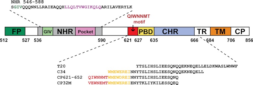
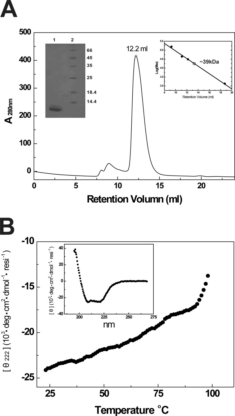
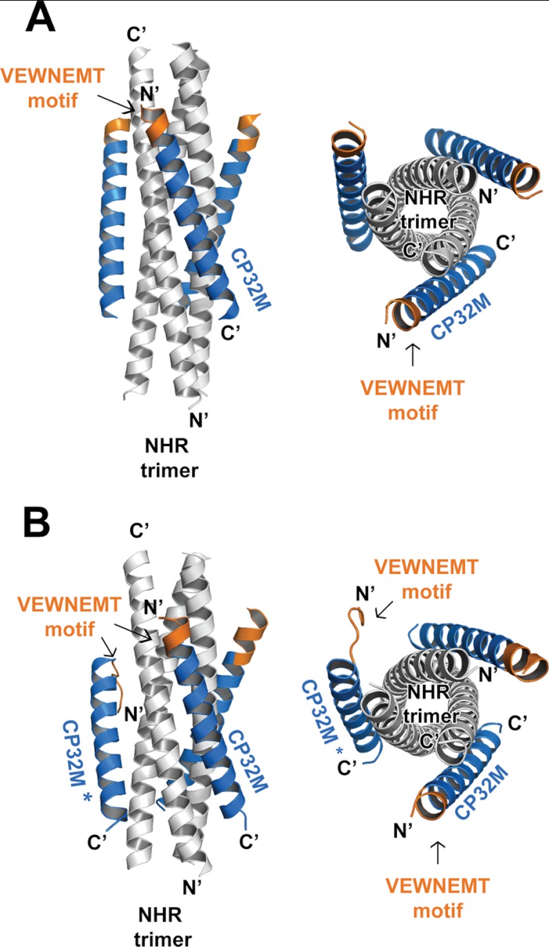
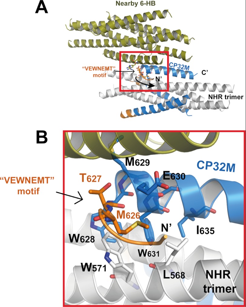
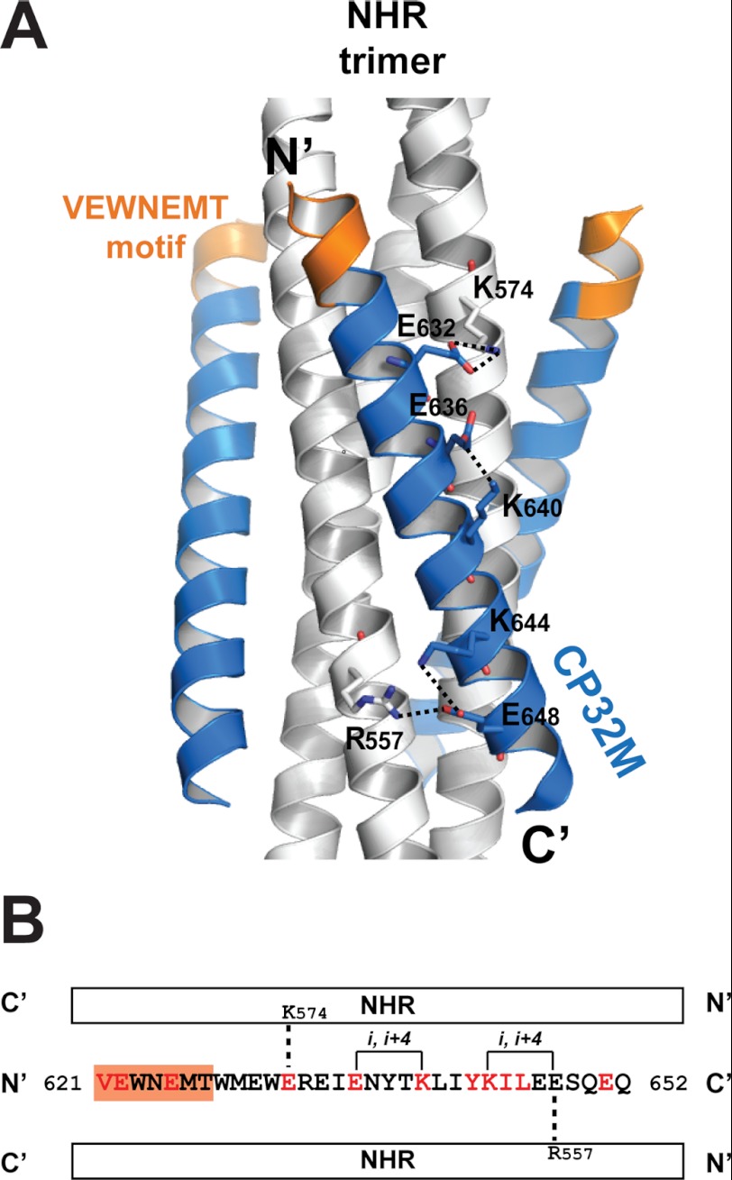
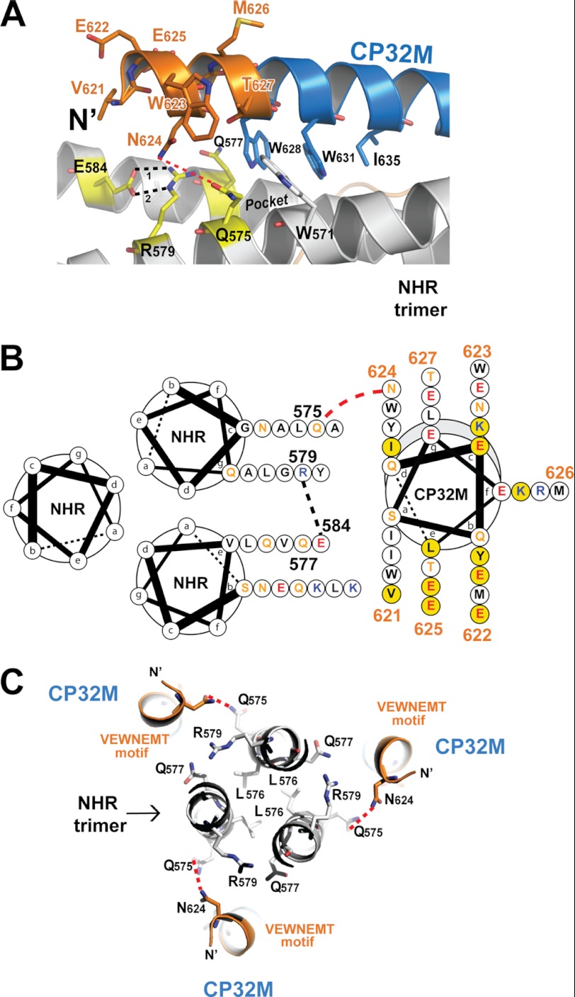
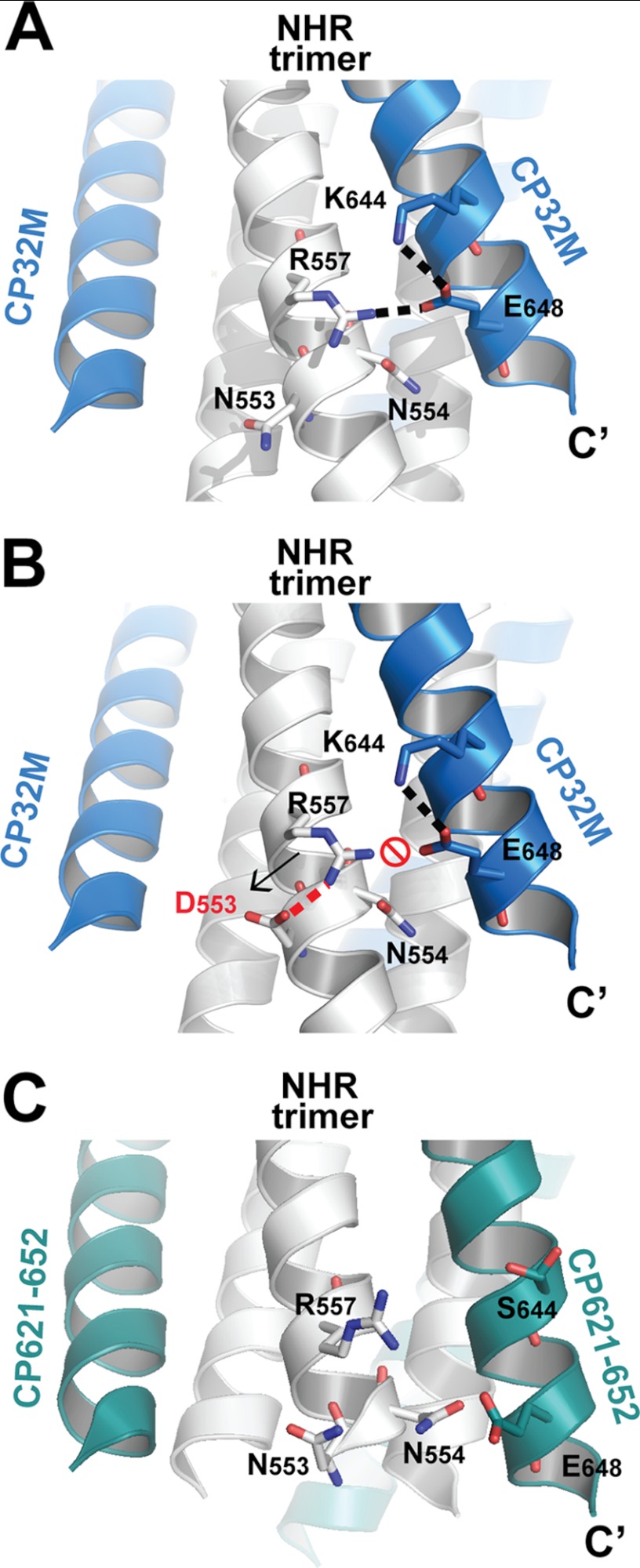
CP32M is a newly designed peptide fusion inhibitor possessing potent anti-HIV activity, especially against T20-resistant HIV-1 strains. In this study, we show that CP32M can efficiently inhibit a large panel of diverse HIV-1 variants, including subtype B', CRF07_BC, and CRF01_AE recombinants and naturally occurring or induced T20-resistant viruses. To elucidate its mechanism of action, we determined the crystal structure of CP32M complexed with its target sequence. Differing from its parental peptide, CP621-652, the (621)VEWNEMT(627) motif of CP32M folds into two α-helix turns at the N terminus of the pocket-binding domain, forming a novel layer in the six-helix bundle structure. Prominently, the residue Asn-624 of the (621)VEWNEMT(627) motif is engaged in the polar interaction with a hydrophilic ridge that borders the hydrophobic pocket on the N-terminal coiled coil. The original inhibitor design of CP32M provides several intra- and salt bridge/hydrogen bond interactions favoring the stability of the helical conformation of CP32M and its interactions with N-terminal heptad repeat (NHR) targets. We identified a novel salt bridge between Arg-557 on the NHR and Glu-648 of CP32M that is critical for the binding of CP32M and resistance against the inhibitor. Therefore, our data present important information for developing novel HIV-1 fusion inhibitors for clinical use.


Figures

FIGURE 1.
FIGURE 1.
Schematic illustration of HIV-1 gp41 functional regions and NHR- or CHR-derived peptide sequences. Shown are the residue numbers of each region corresponding to their positions in gp160 of HIV-1HXB2. FP, fusion peptide; TM, transmembrane domain; TR, tryptophan-rich domain; CP, cytoplasmic domain. The residues corresponding to the NHR pocket region are marked in magenta; the residues for the PBD are marked in yellow; the critical motif adjacent to the N-terminal of PBD is marked in red; the hot spot GIV motif for drug resistance on NHR is marked in green.
FIGURE 2.
FIGURE 2.
Assembly and biophysical characterizations of the 6-HB formed by gp41 NHR546–588/CP32M chimera. A, size-exclusion chromatography and SDS-PAGE analyses of HIV gp41 NHR546–588/CP32M chimera. Chromatographic profile shows UV absorbance at 280 nm. Upper right insert, the log (molecular weight) values of the standard proteins for size-exclusion column calibration (Superdex 75 10/300 GL) are plotted as the function of Ve (elution volume) (●). The data are fitted linearly to derive the standard curve. The molecular mass of HIV gp41 NHR546–588/CP32M chimera is calculated as∼39 kDa (○). Upper left insert, SDS-PAGE analysis of purified HIV gp41 NHR546–588/CP32M chimera. Lane 1, HIV gp41 NHR546–588/CP32M chimera (∼12kDa); lane 2, molecular weight standards. B, the melting curve of NHR546–588/CP32M chimera showing the thermostability of NHR546–588/CP32M chimera. Insert, normalized circular dichroism spectrum showing the α-helical conformation of the 6-HB formed by the chimera. resi, the number of the residues.
FIGURE 3.
FIGURE 3.
Overall structure of the 6-HB structures formed by HIV gp41 NHR546–588/CP32M chimera. Ribbon models of two 6-HB structures formed by NHR546–588/CP32M chimera. The NHR trimers are colored in gray, the CP32M peptides are colored in blue, and the VEWNEMT motifs are colored in orange and labeled. The same color scheme as for the 6-HB is used in all of the figures that follow (Figs. 4–7). The CP32M molecule with the disordered N-terminal motif is marked with an asterisk. A, crystal form 1 (space group P321). Left, side view; right, top view. B, crystal form 2 (space group P21). Left, side view; right, top view.
FIGURE 4.
FIGURE 4.
Crystal packing interactions caused the conformational rearrangement of the N-terminal motif of one particular CP32M in crystal form 2. A, ribbon model of a 6-HB formed by the NHR546–588/CP32M chimera (positioned horizontally) collides with a nearby 6-HB (colored in olive), causing the conformational rearrangement of the N-terminal VEWNEMT motif of one particular CP32M. The collision site is highlighted with a red box. B, magnification of the red-boxed area in A. The VEWNEMT motif of the CP32M peptide is largely disordered, whereas the conserved residues Met-626 and Thr-627 adopt the M-T hook-like structure similar to that found in the crystal structure of CP621–625 (26). The M-T hook residues and the residues interacting with the M-T hook are shown in stick model with labels.
FIGURE 5.
FIGURE 5.
The molecular determinants underlying the stability of the helical conformation of CP32M and the 6-HB structure. A, ribbon model of the 6-HB formed by NHR546–588/CP32M chimera with the charged residues forming the intra- and interhelical salt bridges/hydrogen bonds shown in stick model with labels. The salt bridges/hydrogen bonds are indicated by dashed lines. B, the sequence of CP32M with various features indicated. A single CP32M peptide interacts with two NHR helices (white bars). The residues changed during the inhibitor design are colored in red; the unchanged residues are colored in black. The VEWNEMT motif is highlighted by an orange background. The intrahelical salt bridges/hydrogen bonds observed in the crystal structures are indicated by solid lines; the interhelical salt bridges/hydrogen bonds observed in the crystal structures are indicated by dashed lines.
FIGURE 6.
FIGURE 6.
The VEWNEMT motif of CP32M targets a hydrophilic region on the NHR trimer and form a novel layer of the 6-HB structure. A, a portion of a ribbon model of the 6-HB structure formed by the NHR546–588/CP32M chimera (positioned horizontally). The residues on the N-terminal VEWNEMT motif of CP32M, the residues of the pocket-binding domain, and the residues of the NHR binding site of CP32M are shown in stick model with labels. The hydrophilic ridge comprising Gln-575, Gln-577, Glu-584, and Arg-579 is colored in yellow. The hydrophobic pocket on NHR is indicated. Hydrogen bonds 1 and 2 between Glu-584 and Arg-579 are indicated by black dashed lines. The possible hydrogen bond between Asn-624 and Gln-575 is indicated by a red dashed line. B, helical wheel presentation showing the interaction between CP32M and the NHR trimer. Three NHR helices and one CP32M helix are shown as helical wheel projections. The view is from the bottom of the 6-HB. Three NHR helices form the central coiled coil, and a CP32M helix is packed against the interhelical groove between two NHR helices. At the top of the complex, the N terminus of CP32M (gray shading) is slightly tilted toward the upper NHR helix, and at the bottom of the complex, the C-terminal of the CP32M is tilted toward the lower NHR helix. The color code for the residues is: black, hydrophobic; orange, uncharged; blue, positively charged; red, negatively charged. Residues on the VEWNEMT motif of CP32M are labeled with the residue numbers. Residues mutated from the parental sequence to generate CP32M are highlighted by a yellow background. Red dashed lines indicate the possible hydrogen bonds between Asn-624 on CP32M and Gln-575 on the NHR helices. Black dashed lines indicate the hydrogen bond between Glu-584 and Arg-579. C, the VEWNEMT motif of CP32M forms the novel layer of the 6-HB. Three Leu-576 residues form the hydrophobic core of the NHR trimer that is locked by three Glu-584–Arg-579 “hasps” (the side chain of Glu-584 comes from the front layer and thus is not visible here; see A). Gln-575, Gln-577, Glu-584, and Arg-579 form a hydrophilic region on the NHR trimer, which mediates the hydrophilic interaction with Asn-624 on the VEWNEMT motif of CP32M. The possible hydrogen bonds between Asn-624 and Gln-575 are indicated by red dashed lines.
FIGURE 7.
FIGURE 7.
Molecular basis of a key residue critical for CP32M resistance. A, a portion of a ribbon model of the 6-HB structure formed by the NHR546–588/CP32M chimera. Residues that are important to CP32M resistance and the interaction with NHR helix are shown in stick model. Glu-648 and Lys-644 form an intrahelical salt bridge on CP32M, and Glu-648 forms another interhelical salt bridge with Arg-557 on the NHR helix. The salt bridges are indicated by a dashed line. B, model (generated by Coot and PyMOL) of the 6-HB structure formed by the NHR546–588/CP32M chimera with mutation N553D (equivalent to N42D). The assumed salt bridge between Asp-553 and Arg-557 (indicated by a red dashed line) will neutralize the positive charge of Arg-557, thus disrupting the interhelical salt bridge between Arg-557 and Glu-648 on CP32M and disfavoring the electrostatic interaction between CP32M and its NHR target. C, a portion of a ribbon model of the 6-HB structure formed by T21/CP621–652 peptide (Protein Data Bank ID 3VGX). The NHR helices are colored in gray, and CP621–652 peptide is colored in green. The corresponding residues shown in A and B are shown here in stick model with labels. The corresponding salt bridge between Arg-557 and Glu-648 (distance 4.8 Å) cannot be identified in this structure.

Similar articles

Cited by

References

    1. Colman P. M., Lawrence M. C. (2003) The structural biology of type I viral membrane fusion. Nat. Rev. Mol. Cell Biol. 4, 309–319 - PubMed
    1. Zhu P., Liu J., Bess J., Jr., Chertova E., Lifson J. D., Grisé H., Ofek G. A., Taylor K. A., Roux K. H. (2006) Distribution and three-dimensional structure of AIDS virus envelope spikes. Nature 441, 847–852 - PubMed
    1. Harrison S. C. (2008) Viral membrane fusion. Nat. Struct. Mol. Biol. 15, 690–698 - PMC - PubMed
    1. Eckert D. M., Kim P. S. (2001) Mechanisms of viral membrane fusion and its inhibition. Annu. Rev. Biochem. 70, 777–810 - PubMed
    1. Chan D. C., Kim P. S. (1998) HIV entry and its inhibition. Cell 93, 681–684 - PubMed

Publication types

LinkOut - more resources