

Case Reports
doi: 10.1136/bcr.07.2011.4487.
Two cases of massive mitral annular calcification mimicking left atrial neoplasms
Affiliations
- PMID: 22679051
- PMCID: PMC3176365
- DOI: 10.1136/bcr.07.2011.4487
Item in Clipboard
Case Reports
Two cases of massive mitral annular calcification mimicking left atrial neoplasms
BMJ Case Rep.
.
Abstract
The authors describe two cases of massive mitral annular calcification, initially picked up on echocardiography and suspected of being neoplastic. Subsequent evaluation by CT scanning confirmed the location, aetiology, structure and diagnosis. Both cases demonstrated large masses, with calcification of varying density. This is likely explained by the presence of the previously reported amorphous caseous material demonstrated to be present within such mass structures. Such a feature is described as caseous degeneration. Both patients described have been managed conservatively with medical therapy, predominantly due to their age and general frailty.
Conflict of interest statement
Figures
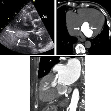
Echocardiogram in left parasternal view (a) shows a mass in the posterior mitral region. A precontrast axial CT short image (b) shows a large highly calcified mass with the long-axis (c) contrast image showing its precise location (arrows) and variable density (high as large arrow and low as black star).

Chest x-ray (a) shows dense calcification in the mitral area (arrows) which on a magnified echocardiographic four chamber view (b) is demonstrated to be an ellipsoid mass (arrows). A contrast short-axis CT scan (c) shows the location of the large calcified mass (arrows) in the region of posterior mitral annulus, outlined on the inside by the posterior mitral leaflet (small arrows) and containing high and low density regions.
Similar articles
-
Caseous Mitral Annular Calcification (CMAC) in an octogenarian with Calcific LV Aneurysm.Int J Cardiovasc Imaging. 2020 Jul;36(7):1291-1292. doi: 10.1007/s10554-020-01827-3. Epub 2020 Mar 30. Int J Cardiovasc Imaging. 2020. PMID: 32232624
-
Caseous calcification of the mitral annulus mimicking benign cardiac tumour of the mitral valve.Cardiovasc J Afr. 2021 Jul-Aug 23;32(4):224-227. doi: 10.5830/CVJA-2021-007. Epub 2021 May 7. Cardiovasc J Afr. 2021. PMID: 34128949 Free PMC article.
-
Severe mitral regurgitation and heart failure due to caseous calcification of the mitral annulus.Cardiology. 2011;118(2):79-82. doi: 10.1159/000326850. Epub 2011 Apr 20. Cardiology. 2011. PMID: 21508639
-
[Caseous calcification of the mitral annulus: case report and review of the literature].G Ital Cardiol (Rome). 2022 Nov;23(11):872-875. doi: 10.1714/3900.38826. G Ital Cardiol (Rome). 2022. PMID: 36300390 Review. Italian.
-
Calcified Amorphous Tumor Causing Shower Embolism to the Brain: A Case Report with Serial Echocardiographic and Neuroradiologic Images and a Review of the Literature.J Stroke Cerebrovasc Dis. 2017 May;26(5):e85-e89. doi: 10.1016/j.jstrokecerebrovasdis.2017.02.019. Epub 2017 Mar 18. J Stroke Cerebrovasc Dis. 2017. PMID: 28318955 Review.
Cited by
-
Imaging of cardiac valves by computed tomography.Scientifica (Cairo). 2013;2013:270579. doi: 10.1155/2013/270579. Epub 2013 Dec 29. Scientifica (Cairo). 2013. PMID: 24490107 Free PMC article.
-
Surgical removal of calcified amorphous tumor localized to mitral valve leaflet without mitral annular calcification.Surg Case Rep. 2015 Dec;1(1):39. doi: 10.1186/s40792-015-0040-6. Epub 2015 May 1. Surg Case Rep. 2015. PMID: 26943404 Free PMC article.
References
-
- Harpaz D, Auerbach I, Vered Z, et al. Caseous calcification of the mitral annulus: a neglected, unrecognized diagnosis. J Am Soc Echocardiogr 2001;14:825–31 - PubMed
-
- Fox CS, Vasan RS, Parise H, et al. ; Framingham Heart Study Mitral annular calcification predicts cardiovascular morbidity and mortality: the Framingham Heart Study. Circulation 2003;107:1492–6 - PubMed
-
- Barasch E, Gottdiener JS, Larsen EK, et al. Clinical significance of calcification of the fibrous skeleton of the heart and aortosclerosis in community dwelling elderly. The cardiovascular health study (CHS). Am Heart J 2006;151:39–47 - PubMed
-
- Deluca G, Correale M, Ieva R, et al. The incidence and clinical course of caseous calcification of the mitral annulus: a prospective echocardiographic study. J Am Soc Echocardiogr 2008;21:828–33 - PubMed
Publication types
MeSH terms
LinkOut - more resources
Full Text Sources
Medical