

Cardiac micro-computed tomography imaging of the aging coronary vasculature
- PMID: 22679058
- PMCID: PMC3408091
- DOI: 10.1161/CIRCIMAGING.112.973057
Cardiac micro-computed tomography imaging of the aging coronary vasculature
Abstract
Background: Alterations at the level of the coronary circulation with aging may play an important role in the evolution of age-associated changes in left ventricular (LV) fibrosis and function. However these age-associated changes in the coronary vasculature remain poorly defined primarily due to the lack of high resolution imaging technologies. The current study was designed to utilize cardiac micro-computed tomography (micro-CT) technology as a novel imaging strategy, to define the 3-dimensional coronary circulation in the young and aged heart and its relationship to LV fibrosis and function.
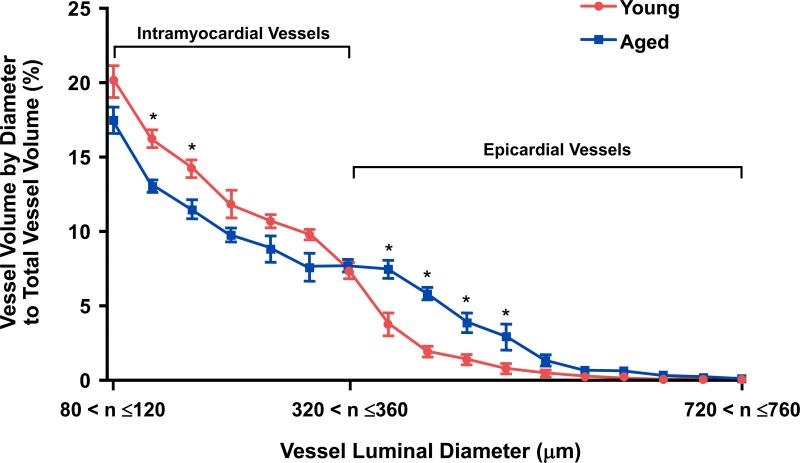
Methods and results: Young (2 months old; n=10) and aged (20 months old; n=10) Fischer rats underwent cardiac micro-CT imaging as well as echocardiography, blood pressure, and fibrosis analysis. Importantly, when indexed to LV mass, which increased with age, the total and intramyocardial vessel volumes were lower, whereas the epicardial vessel volume, with and without indexing to LV mass, was significantly higher in the aged hearts compared with the young hearts. Moreover, the aged hearts had a significantly lower percentage of intramyocardial vessel volume and a significantly higher percentage of epicardial vessel volume, when normalized to the total vessel volume, compared with the young hearts. Further, the aged hearts had significant LV fibrosis and mild LV dysfunction compared with the young hearts.
Conclusions: This micro-CT imaging study reports the reduction in normalized intramyocardial vessel volume within the aged heart, in association with increased epicardial vessel volume, in the setting of increased LV fibrosis, and mild LV dysfunction.
Figures


Similar articles
-
Age-associated changes in hearts of male Fischer 344/Brown Norway F1 rats.Ann Clin Lab Sci. 2006 Autumn;36(4):427-38. Ann Clin Lab Sci. 2006. PMID: 17127729
-
New Parameter Derived from Three-Dimensional Speckle-Tracking Echocardiography for the Estimation of Left Ventricular Filling Pressure in Nondilated Hearts.J Am Soc Echocardiogr. 2017 May;30(5):522-531. doi: 10.1016/j.echo.2017.01.015. Epub 2017 Mar 18. J Am Soc Echocardiogr. 2017. PMID: 28325672
-
Cardiac dysfunction in aging conscious rats: altered cardiac cytoskeletal proteins as a potential mechanism.Am J Physiol Heart Circ Physiol. 2008 Aug;295(2):H860-6. doi: 10.1152/ajpheart.00146.2008. Epub 2008 Jun 20. Am J Physiol Heart Circ Physiol. 2008. PMID: 18567712 Free PMC article.
-
Assessment of left sided filling dynamics in diastolic dysfunction using cardiac computed tomography.Eur J Radiol. 2015 Oct;84(10):1930-7. doi: 10.1016/j.ejrad.2015.07.006. Epub 2015 Jul 13. Eur J Radiol. 2015. PMID: 26205972
-
Residual fibrosis affects a long-term result of left ventricular volume reduction surgery for dilated cardiomyopathy in a rat experimental study.Eur J Cardiothorac Surg. 2004 Dec;26(6):1174-9. doi: 10.1016/j.ejcts.2004.06.023. Eur J Cardiothorac Surg. 2004. PMID: 15541980
Cited by
-
A Review of Ex Vivo X-ray Microfocus Computed Tomography-Based Characterization of the Cardiovascular System.Int J Mol Sci. 2021 Mar 23;22(6):3263. doi: 10.3390/ijms22063263. Int J Mol Sci. 2021. PMID: 33806852 Free PMC article. Review.
-
μCT imaging of a multi-organ vascular fingerprint in rats.PLoS One. 2024 Oct 14;19(10):e0308601. doi: 10.1371/journal.pone.0308601. eCollection 2024. PLoS One. 2024. PMID: 39401231 Free PMC article.
-
Correlative Imaging of the Murine Hind Limb Vasculature and Muscle Tissue by MicroCT and Light Microscopy.Sci Rep. 2017 Feb 7;7:41842. doi: 10.1038/srep41842. Sci Rep. 2017. PMID: 28169309 Free PMC article.
-
Microcomputed analysis of nerve angioarchitecture after combined stem cell delivery and surgical angiogenesis to nerve allograft.J Plast Reconstr Aesthet Surg. 2021 Aug;74(8):1919-1930. doi: 10.1016/j.bjps.2020.12.039. Epub 2020 Dec 24. J Plast Reconstr Aesthet Surg. 2021. PMID: 33436338 Free PMC article.
-
Micro-CT of rodents: state-of-the-art and future perspectives.Phys Med. 2014 Sep;30(6):619-34. doi: 10.1016/j.ejmp.2014.05.011. Epub 2014 Jun 26. Phys Med. 2014. PMID: 24974176 Free PMC article. Review.
References
-
- Olivetti G, Melissari M, Capasso JM, Anversa P. Cardiomyopathy of the aging human heart. Myocyte loss and reactive cellular hypertrophy. Circ Res. 1991;68:1560–1568. - PubMed
-
- Anversa P, Capasso JM. Cellular basis of aging in the mammalian heart. Scanning Microscopy. 1991;5:1065–1073. - PubMed
-
- Weber KT, Brilla CG. Pathological hypertrophy and cardiac interstitium. Fibrosis and renin-angiotensin-aldosterone system. Circulation. 1991;83:1849–1865. - PubMed
-
- Lakatta EG, Levy D. Arterial and cardiac aging: major shareholders in cardiovascular disease enterprises: Part II: the aging heart in health: links to heart disease. Circulation. 2003;107:346–354. - PubMed
Publication types
MeSH terms
Grants and funding
LinkOut - more resources
Full Text Sources
Medical
