. 2012 Jun 8;336(6086):1310-4.
doi: 10.1126/science.1221708.

Actin network architecture can determine myosin motor activity

Affiliations

Actin network architecture can determine myosin motor activity

Anne-Cécile Reymann et al. Science. .

Abstract

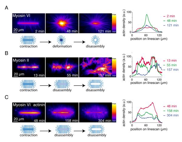
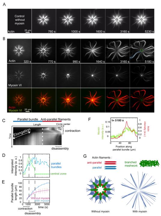
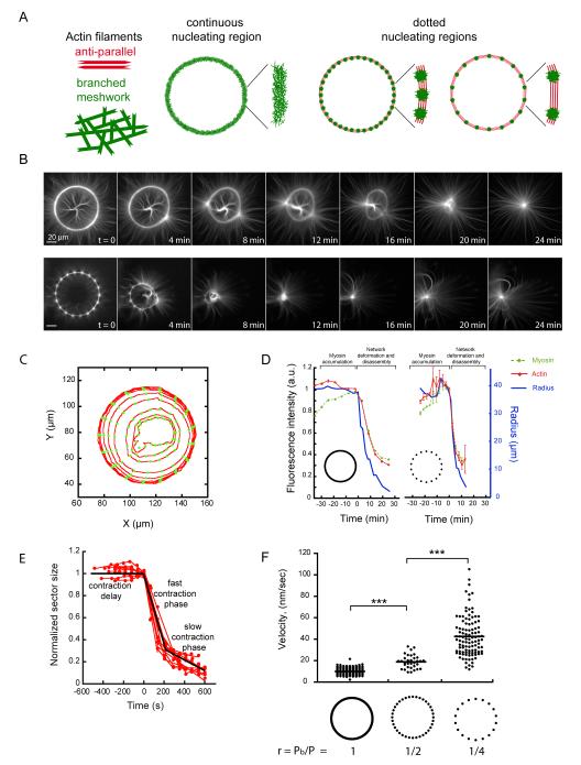
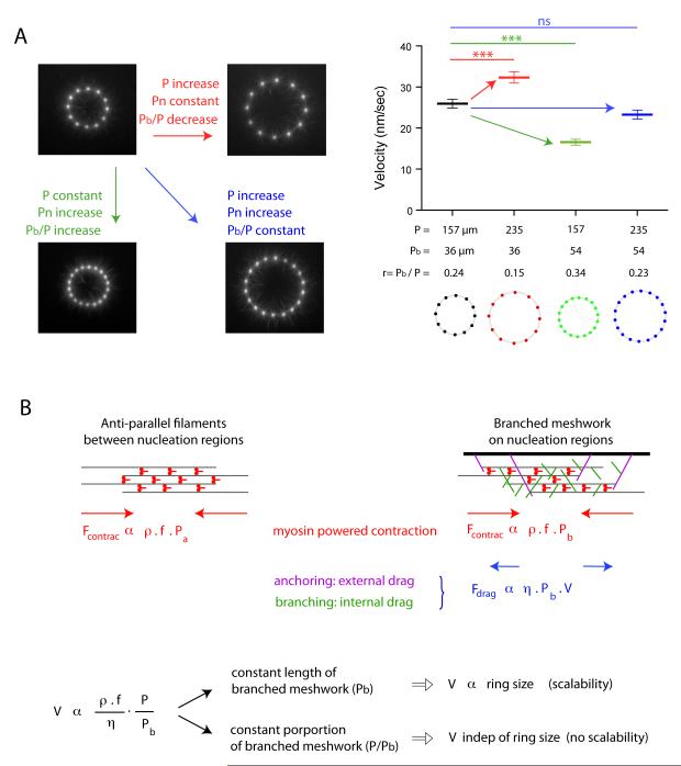
The organization of actin filaments into higher-ordered structures governs eukaryotic cell shape and movement. Global actin network size and architecture are maintained in a dynamic steady state through regulated assembly and disassembly. Here, we used experimentally defined actin structures in vitro to investigate how the activity of myosin motors depends on network architecture. Direct visualization of filaments revealed myosin-induced actin network deformation. During this reorganization, myosins selectively contracted and disassembled antiparallel actin structures, while parallel actin bundles remained unaffected. The local distribution of nucleation sites and the resulting orientation of actin filaments appeared to regulate the scalability of the contraction process. This "orientation selection" mechanism for selective contraction and disassembly suggests how the dynamics of the cellular actin cytoskeleton can be spatially controlled by actomyosin contractility.


Figures

Fig. 1
Fig. 1
Myosin induced actin meshwork contraction and disassembly (A) Time-series of myosin VI-induced network contraction on a bar-shaped micropattern. Actin filaments were visualized with fluorescent monomers. “Fire” look-up table color-coding reveals variations in actin network densities, quantified with a linescan along the bar at different time points. Actin density peaks because of network deformation after 48 minutes then falls off because of network disassembly. (B) Same as (A) with muscle myosin II-induced contraction. (C) Same as (A) with 100 nM alpha-actinin in the polymerization mix.
Fig. 2
Fig. 2
Regioselective action of myosins (A) Time-series of network assembly on an eight-branch actin nucleating radial array. (B) Time-series of myosin VI-induced architecture selective contraction and disassembly (actin, myosin and an overlay are shown). (C) Kymograph of actin fluorescence along a parallel bundle (blue dashed line in B) 5180 s) and central region of actin filaments (dashed green circle in B) 5180 s), showing the different localization of elongation and contraction, disassembly. (D) Fluorescence intensity of a central zone (dashed green circle in B) and a parallel bundle (blue dashed line in B) over time. (E) Length variations of parallel bundles over time in absence or presence of myosins. (F) Linescan of fluorescence intensity along a parallel bundle confirming myosin presence all along. (G) Schematic representation of the final architecture on an eight-branch actin nucleating radial array in absence and in presence of myosins in solution.
Fig. 3
Fig. 3
The proportion of anti-parallel filaments regulates network contraction rate. (A) Schematic representation of actin networks nucleated on a full and dotted rings. (B) Time-series of myosin-induced contraction of actin networks nucleated from full (top) and dotted (bottom) rings. (C) Illustration of automated network contraction analysis (23). Each circle represents a time point. (D) The radius and total fluorescence intensities of both actin and myosin were recorded for all angular sectors over time. (E) Ring constriction kinetics. Time series of length values (red dots) could be fitted by three distinct phases (black line). (F) Fast contraction phase velocity measurements were compared between various ring compositions.
Fig. 4
Fig. 4
The proportion of branched meshwork regulates the scalability of ring contraction. (A) Respective effects of size and proportion of branched meshwork in contraction kinetics. We varied the ring perimeter P and the length of that perimeter nucleating a branched meshwork Pbranched (Pb) independently. Images show an early time point during actin network assembly on micropatterned dots. Fast contraction phase velocity measurements were compared between various ring configurations. (B) Model description. Filaments assemble into anti-parallel bundles between nucleation regions (left scheme). Nucleation regions (wide black bar, right scheme) generate branched actin meshwork. The contraction force is proportional to the density of myosins per unit length of filament, ρ, to the force per myosin head, f, and to the portion of the perimeter made of the relevant network, Pa for the antiparallel bundles and Pb for the branched meshwork. Myosin density is constant over the entire perimeter P=Pa+Pb. Anti-parallel bundles have a friction drag negligible compared to branched meshwork in which the effective friction coefficient η has two origins: an external drag due to network anchoring on the nucleation region and an internal drag due to entanglement of filament branches. The balance between the total contraction force and the frictional drag sets the contraction velocity V, which appeared to be proportional to the ratio P/Pb as observed in all our experiments.

References

    1. Cramer LP, Mitchison TJ. Myosin is involved in post-mitotic cell spreading. J. Cell Biol. 1995;131:1. - PMC - PubMed
    1. Vignjevic D, et al. Formation of filopodia-like bundles in vitro from a dendritic network. J. Cell Biol. 2003;160:951. - PMC - PubMed
    1. Cramer LP, Siebert M, Mitchison TJ. Identification of novel graded polarity actin filament bundles in locomoting heart fibroblasts: implications for the generation of motile force. J. Cell Biol. 1997;136:1287. - PMC - PubMed
    1. Verkhovsky AB, Svitkina TM, Borisy GG. Myosin II filament assemblies in the active lamella of fibroblasts: their morphogenesis and role in the formation of actin filament bundles. J. Cell Biol. 1995;131:989. - PMC - PubMed
    1. Svitkina TM, Verkhovsky AB, McQuade KM, Borisy GG. Analysis of the actin-myosin II system in fish epidermal keratocytes: mechanism of cell body translocation. J. Cell Biol. 1997;139:397. - PMC - PubMed

Publication types