Review
. 2012 Aug;14(8):979-93.
doi: 10.1093/neuonc/nos135. Epub 2012 Jun 7.

In vivo models of primary brain tumors: pitfalls and perspectives

Affiliations
Review

In vivo models of primary brain tumors: pitfalls and perspectives

Peter C Huszthy et al. Neuro Oncol. 2012 Aug.

Abstract

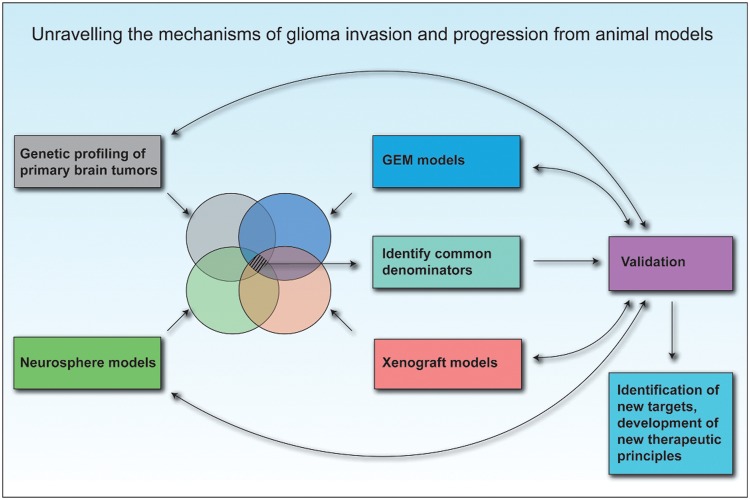
Animal modeling for primary brain tumors has undergone constant development over the last 60 years, and significant improvements have been made recently with the establishment of highly invasive glioblastoma models. In this review we discuss the advantages and pitfalls of model development, focusing on chemically induced models, various xenogeneic grafts of human cell lines, including stem cell-like cell lines and biopsy spheroids. We then discuss the development of numerous genetically engineered models available to study mechanisms of tumor initiation and progression. At present it is clear that none of the current animal models fully reflects human gliomas. Yet, the various model systems have provided important insight into specific mechanisms of tumor development. In particular, it is anticipated that a combined comprehensive knowledge of the various models currently available will provide important new knowledge on target identification and the validation and development of new therapeutic strategies.


Figures

Fig. 1.
Fig. 1.
Key events in brain tumor modeling in animals. Milestones in brain tumor model development starting with transplantation of human xenografts into immunocompetent rodents in the 1940s through rodent carcinogenesis and human monolayer cell line development in the 1960s, toward the establishment of GEM models in the 1990s, and finally the establishment of xenograft models based on stem cell enrichment.
Fig. 2.
Fig. 2.
Histological features of chemically induced gliosarcoma models. (A and B) Collective infiltration of N-ethylnitrosourea–induced BT4C cells into the brain tissue (frequently referred to as mesenchymal chain movement). Black arrows indicate the leading edge of migrating tumor cells. (A) Green arrows mark peritumoral necrosis and microhemorrhages. (C) Growth pattern of BT4C cells in the tumor bed. (D) Region of a sarcomatous growth with elongated, spindle-shaped cells and extracellular matrix production. All scalebars are 20 µm, except A, which is 50 µm.
Fig. 3.
Fig. 3.
Histological features of GBM xenograft models. (A) GBM patient biopsies may be processed to yield adherent cell lines in serum-containing medium, which results in an extensive clonal selection and cellular adaptation process. Xenografts generated from such cell lines will display angiogenic growth and well-defined borders toward the brain tissue. No single tumor-cell invasion is seen (the example here is from the U-87 glioma cell line). (B) Enzymatic dissociation of patient biopsies with subsequent culture in neurobasal serum-free medium selects for a highly tumorigenic subpopulation in human GBM. In several instances, the resulting xenografts are highly infiltrative, following white matter tracts and spreading over the corpus callosum. (C) The biopsy xenograft model maintains several tumor cell clones from the biopsy, as well as other cell types and extracellular matrix components. When passaged extensively in immunodeficient animals, the xenografts maintain their invasive growth and develop other characteristics of human GBM, such as dilated vessels, angiogenesis, and pseudopalisading necrosis. Scale bars: 100 µm.
Fig. 4.
Fig. 4.
Strategy for separation and analysis of the tumor/host cellular compartments. With the development of immunodeficient mice expressing enhanced green fluorescent protein, it is at present possible to completely separate and immunophenotype the cells in the tumor/host cellular compartments.137 This technique shows considerable promise in elucidating mechanisms involved in tumor/host cell communication.
Fig. 5.
Fig. 5.
Unraveling the mechanisms of tumor invasion and progression from animal models. Further development in the field of neuro-oncology must integrate current molecular knowledge from human tumors as well as biological knowledge from GEM and spheroid and stem/progenitor cell–based xenotransplantation models. The search for common denominators will most likely lead to the identification of robust therapeutic targets that should eventually be validated in human GBM.

Similar articles

Cited by

References

    1. Kerbel RS. What is the optimal rodent model for anti-tumor drug testing? Cancer Metastasis Rev. 1998;17(3):301–304. - PubMed
    1. Kerbel RS. Human tumor xenografts as predictive preclinical models for anticancer drug activity in humans: better than commonly perceived—but they can be improved. Cancer Biol Ther. 2003;2(4 suppl 1):S134–S139. - PubMed
    1. Peterson JK, Houghton PJ. Integrating pharmacology and in vivo cancer models in preclinical and clinical drug development. Eur J Cancer. 2004;40(6):837–844. - PubMed
    1. Hesselager G, Holland EC. Using mice to decipher the molecular genetics of brain tumors. Neurosurgery. 2003;53(3):685–694. discussion 695. - PubMed
    1. Hanahan D, Weinberg RA. The hallmarks of cancer. Cell. 2000;100(1):57–70. - PubMed

Publication types