Chronic flank pain, fever and an unusual diagnosis
- PMID: 22679166
- PMCID: PMC3189649
- DOI: 10.1136/bcr.08.2011.4646
Chronic flank pain, fever and an unusual diagnosis
Abstract
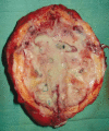
Xanthogranulomatous pyelonephritis (XGP) is a rare, serious, debilitating illness characterised by an infectious renal phlegmon. Most cases of XGP are unilateral and are often associated with urinary tract obstruction, infection, nephrolithiasis, diabetes, and/or immune compromise. This disease process ultimately results in focal or diffuse renal destruction and is characterised pathologically by lipid-laden foamy macrophages. XGP occurs in approximately 1% of all renal infections. The kidney is usually non-functional. XGP displays neoplasm like properties capable of local tissue invasion and destruction and has been referred to as a pseudotumour. Adjacent organs including the spleen, pancreas or duodenum may be involved. The gross appearance of XGP is a mass of yellow tissue with regional necrosis and haemorrhage, superficially resembling renal cell carcinoma. Renal cell carcinoma may be indistinguishable from XGP radiographically and clinically. The treatment of XGP is almost universally extirpative and can pose a formidable challenge to the surgeon.
Conflict of interest statement
Figures


References
-
- Mishriki YY, Doehne K. Puzzles in practice. Xanthogranulomatous pyelonephritis (XGP). Postgrad Med 2010;122:230–2 - PubMed
-
- Charrada-Ben Farhat L, Saïed W, Dali N, et al. [Imaging features of xanthogranulomatous pyelonephritis]. J Radiol 2007;88(9 Pt 1):1171–7 - PubMed
-
- Tiguert R, Gheiler EL, Yousif R, et al. Focal xanthogranulomatous pyelonephritis presenting as a renal tumor with vena caval thrombus. J Urol 1998;160:117–18 - PubMed
-
- Shah HN, Jain P, Chibber PJ. Renal tuberculosis simulating xanthogranulomatous pyelonephritis with contagious hepatic involvement. Int J Urol 2006;13:67–8 - PubMed
-
- Hitti W, Drachenberg C, Cooper M, et al. Xanthogranulomatous pyelonephritis in a renal allograft associated with xanthogranulomatous diverticulitis: report of the first case and review of the literature. Nephrol Dial Transplant 2007;22:3344–7 - PubMed
Publication types
MeSH terms
LinkOut - more resources
Full Text Sources
