. 2012 Nov;5(6):823-33.
doi: 10.1242/dmm.008557. Epub 2012 May 31.

Interleukin-1 receptor antagonist is beneficial after subarachnoid haemorrhage in rat by blocking haem-driven inflammatory pathology

Affiliations

Interleukin-1 receptor antagonist is beneficial after subarachnoid haemorrhage in rat by blocking haem-driven inflammatory pathology

Andrew D Greenhalgh et al. Dis Model Mech. 2012 Nov.

Abstract

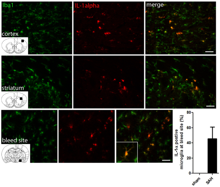
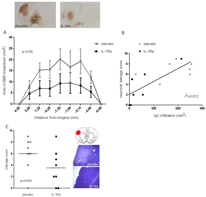
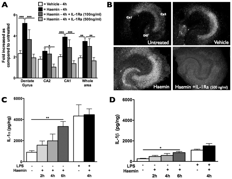
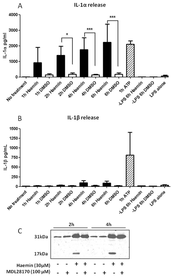
Subarachnoid haemorrhage (SAH) is a major contributor to the burden of stroke on society. Treatment options are limited and animal models of SAH do not always mimic key pathophysiological hallmarks of the disease, thus hindering development of new therapeutics. Inflammation is strongly associated with brain injury after SAH in animals and patients, and inhibition of the pro-inflammatory cytokine interleukin-1 (IL-1) represents a possible therapeutic target. Here we report that a rupture of the middle cerebral artery in the rat produces heterogeneous infarct patterns similar to those observed in human SAH. Administration of the IL-1 receptor antagonist (IL-1Ra) reduced blood-brain barrier breakdown, and the extent of breakdown correlated with brain injury. After SAH, haem oxygenase-1 (HO-1) was strongly expressed around the bleed site and in the cortex and striatum, indicating the presence of free haem, a breakdown product of haemoglobin. HO-1 expression was also found in the same regions as microglial/macrophage expression of IL-1α. The direct effect of haem on IL-1α expression was confirmed in vitro using organotypic slice culture (OSC). Haem-induced cell death was dependent on IL-1 signalling, with IL-1Ra completely blocking cellular injury. Furthermore, stimulation of mouse primary mixed glial cells with haem induced the release of IL-1α, but not IL-1β. Thus, we suggest that haem, released from lysed red blood cells (RBCs) in the subarachnoid space, acts as a danger-associated molecular pattern (DAMP) driving IL-1-dependent inflammation. These data provide new insights into inflammation after SAH-induced brain injury and suggest IL-1Ra as a candidate therapeutic for the disease.


Figures

Fig. 1.
Fig. 1.
IL-1α is expressed by microglia/macrophages early after experimental SAH. Panels show coronal brain sections of SAH animals 12 hours after bleed. Images are representative of the cortex, striatum and adjacent area to the bleed site after SAH. IL-1α-positive fluorescent cells (red); Iba1-positive fluorescent cells show microglia/macrophages (green). Images are merged to reveal IL-1α colocalisation with microglia/macrophages. Graph shows the percentage of IL-1α-positive microglia cells adjacent to the bleed site (n=5). No IL-1α-positive cells were seen in sham controls. Coronal inserts indicate the area that the image was taken from and insert shows high magnification of IL-1α/Iba1 colocalisation. Scale bars: 50 μm.
Fig. 2.
Fig. 2.
Early after SAH, HO-1 is found throughout the brain localised to brain regions expressing IL-1α. Panels show representative coronal brain sections of SAH animals 12 hours after bleed (n=5). Representative images from the area adjacent to the bleed site, the cortex and the striatum are shown. HO-1-positive fluorescent cells (green); IL-1α-positive fluorescent cells (red). Areas of HO-1 immunofluorescence correspond to areas of microglial activation and IL-1α expression seen in Fig. 1. In sham animals, HO-1-positive fluorescence was undetectable. Scale bars: 100 μm.
Fig. 3.
Fig. 3.
Blood load grades 48 hours after SAH. (A) Photomicrographs showing examples of blood load grades 48 hours after SAH. (B) Blood load grades were highly variable between animals; as expected, there was no significant difference between groups that went on to receive IL-1Ra (n=15) or placebo (n=14). (C) Blood load grades ≥7 were eligible for analysis.
Fig. 4.
Fig. 4.
BBB breakdown correlates with neuronal damage and is significantly reduced by IL-1Ra. (A) Volume of BBB breakdown at 48 hours, as measured by IgG infiltration, is reduced by administration of IL-1Ra (s.c. 75 mg/kg) 15 minutes after bleed. Top panels show representative coronal sections from placebo (n=8) and IL-1Ra (n=8) groups. (B) Linear regression analysis shows positive correlation between IgG infiltration and neuronal damage (r2=0.612). (C) Neuronal damage 48 hours after SAH; there is no significant difference between IL-1Ra (n=8) and placebo (n=8) groups (P=0.059). Inserts show representative coronal sections from placebo and IL-1Ra groups and coronal map indicating the area imaged. (A,C) Unpaired Student’s t-tests; P<0.05 considered significant.
Fig. 5.
Fig. 5.
Experimental SAH does not induce inflammatory changes in the periphery or CSF. Log transformed CXCL-1 concentrations in plasma (A) and IL-6 concentrations in CSF (B). Two-way ANOVA showed a significant effect of time after bleed in CXCL-1 plasma concentrations, with no difference between sham or SAH groups. One-way ANOVA showed there was no significant difference in CSF IL-6 concentrations between groups (n=7–10).
Fig. 6.
Fig. 6.
Haemin-induced neuronal death is IL-1 dependent in OSCs. Treatment of OSCs with haemin (30 μM; oxidation product of haem) induced neuronal death, as seen by PI staining, mainly in the dentate gyrus (DG) and CA1 regions of the hippocampus (A). This effect was abolished by co-treatment with IL-1Ra (500 ng/ml). (B) Representative images of PI staining in OSCs after treatment (note that the image intensity of the haemin + IL-1Ra example has been increased in order to visualise the hippocampus). Haemin treatment induced the expression of IL-1α (C) and IL-1β (D), measured by ELISA, with IL-1α being the predominant isoform induced (n=3–5, P=0.0037 between IL-1α and IL-1β after 6 hours). *P<0.05, **P<0.01, ***P<0.001.
Fig. 7.
Fig. 7.
Haemin induces the release of IL-1α but not IL-1β in primary mixed glia. Graphs show the release of IL-1α and IL-1β in LPS-treated mouse primary mixed glial cells after haemin (30 μM; black bars) or vehicle (DMSO; white bars) for 1, 2, 4 and 6 hours. ATP (5 μM) stimulation for 1 hour was used as a positive control (striped bar). IL-1α release was significantly increased after 2, 4 and 6 hours of haem stimulation compared with vehicle (A). Haem stimulation had no effect on release of IL-1β (B). n=4, one-way ANOVA followed by Student’s t-test with Bonferroni’s correction; *P<0.05, **P<0.01, ***P<0.001. Western blot analysis revealed that haemin (30 μM) induced cleavage of pro-IL-1α (31 kDa) to the mature form of IL-1α (17 kDa) through a calpain-1-dependent mechanism, because release of mature IL-1α was blocked by the calpain-1 inhibitor MDL28170 (100 μM) (C).

References

    1. Allan S. M., Tyrrell P. J., Rothwell N. J. (2005). Interleukin-1 and neuronal injury. Nat. Rev. Immunol. 5, 629–640 - PubMed
    1. Ascenzi P., Bocedi A., Visca P., Altruda F., Tolosano E., Beringhelli T., Fasano M. (2005). Hemoglobin and heme scavenging. IUBMB Life 57, 749–759 - PubMed
    1. Borsody M., Burke A., Coplin W., Miller-Lotan R., Levy A. (2006). Haptoglobin and the development of cerebral artery vasospasm after subarachnoid hemorrhage. Neurology 66, 634–640 - PubMed
    1. Brough D., Tyrrell P. J., Allan S. M. (2011). Regulation of interleukin-1 in acute brain injury. Trends Pharmacol. Sci. 32, 617–622 - PubMed
    1. Chaichana K. L., Pradilla G., Huang J., Tamargo R. J. (2010). Role of inflammation (leukocyte-endothelial cell interactions) in vasospasm after subarachnoid hemorrhage. World Neurosurg. 73, 22–41 - PubMed

Publication types

MeSH terms