Clinical Trial
. 2013 May;72(5):665-71.
doi: 10.1136/annrheumdis-2012-201469. Epub 2012 Jun 7.

The ability of synovitis to predict structural damage in rheumatoid arthritis: a comparative study between clinical examination and ultrasound

Affiliations
Clinical Trial

The ability of synovitis to predict structural damage in rheumatoid arthritis: a comparative study between clinical examination and ultrasound

Maxime Dougados et al. Ann Rheum Dis. 2013 May.

Erratum in

  • Ann Rheum Dis. 2013 Jul;72(7):1270. Etcheparre, Frederic [corrected to Etchepare, Fabien]

Abstract

Objectives: To evaluate synovitis (clinical vs ultrasound (US)) to predict structural progression in rheumatoid arthritis (RA).

Methods: Patients with RA.

Study design: Prospective, 2-year follow-up.

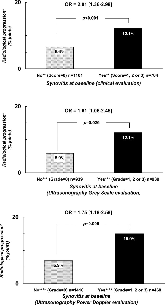
Data collected: Synovitis (32 joints (2 wrists, 10 metacarpophalangeal, 10 proximal interphalangeal, 10 metatarsophalangeal)) at baseline and after 4 months of therapy by clinical, US grey scale (GS-US) and power doppler (PD-US); x-rays at baseline and at year 2.

Analysis: Measures of association (OR) were tested between structural deterioration and the presence of baseline synovitis, or its persistence, after 4 months of therapy using generalised estimating equation analysis.

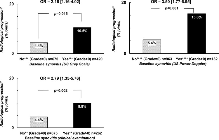
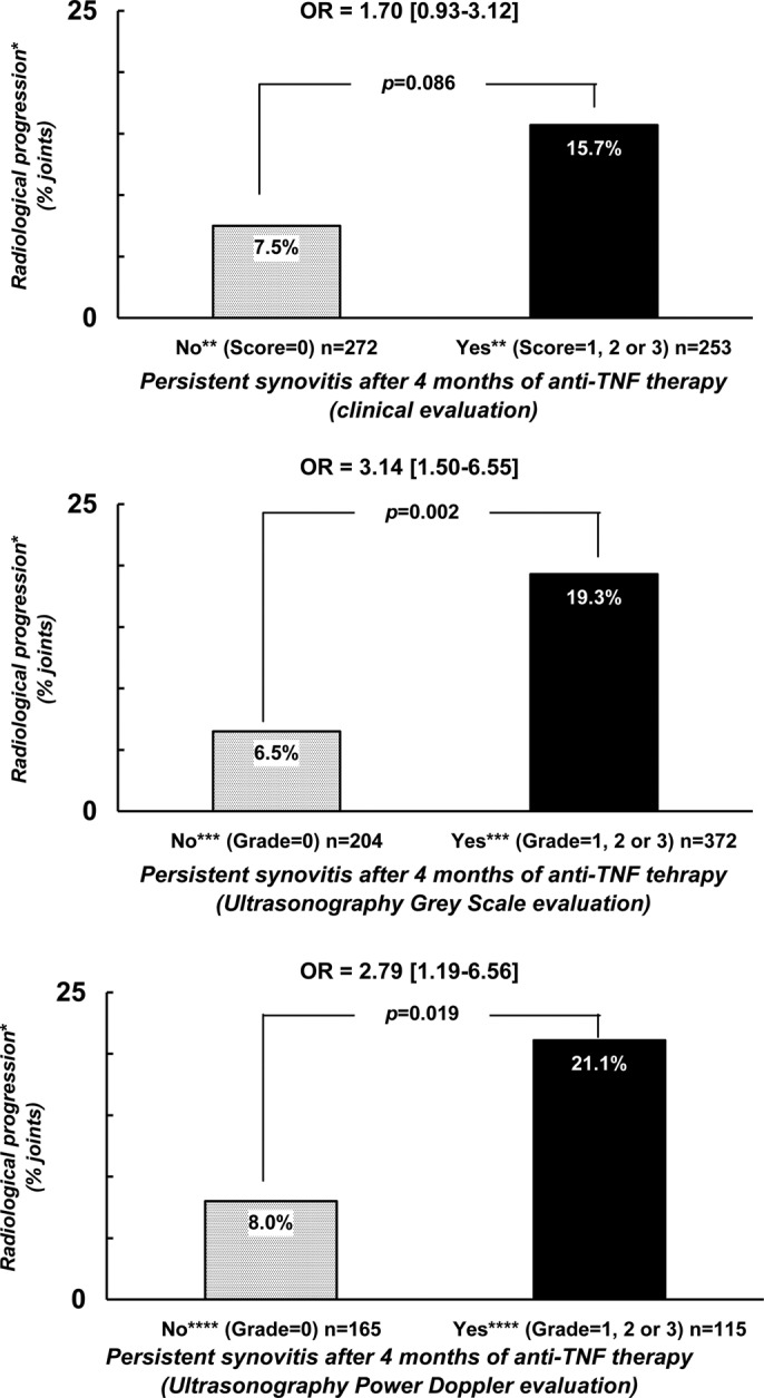
Results: Structural deterioration was observed in 9% of the 1888 evaluated joints in 59 patients. Baseline synovitis increased the risk of structural progression: OR=2.01 (1.36-2.98) p<0.001 versus 1.61 (1.06-2.45) p=0.026 versus 1.75 (1.18-2.58) p=0.005 for the clinical versus US-GS versus US-PD evaluation, respectively. In the joints with normal baseline examination (clinical or US), an increased probability for structural progression in the presence of synovitis for the other modality was also observed (OR=2.16 (1.16-4.02) p=0.015 and 3.50 (1.77-6.95) p<0.001 for US-GS and US-PD and 2.79 (1.35-5.76) p=0.002) for clinical examination. Persistent (vs disappearance) synovitis after 4 months of therapy was also predictive of subsequent structural progression.

Conclusions: This study confirms the validity of synovitis for predicting subsequent structural deterioration irrespective of the modality of examination of joints, but also suggests that both clinical and ultrasonographic examinations may be relevant to optimally evaluate the risk of subsequent structural deterioration.


Conflict of interest statement

Competing interests:

Figures

Figure 1
Figure 1
Probability of radiological progression after a 2-year follow-up period with regard to the presence of baseline synovitis defined by either clinical or ultrasonographic examination. *Radiological progression = occurrence or worsening of erosion or joint space narrowing. **Score of ‘clinical’ synovitis (0 = no synovitis; 1 = doubtful synovitis; 2 = obvious and moderate synovitis; 3 = obvious and important synovitis. ***Score of ultrasonography grey-scale evaluation: 0 = absence of synovial thickening; 1 = mild synovial thickening; 2 = moderate synovial thickening; 3 = marked synovial thickening. ****Score of ultrasonographic Power Doppler evaluation: 0 = absence of signal, no intra-articular flow; 1 = mild, one or two vessels' signals (including one confluent vessel) for small joints and two to three signals for large joints (including two confluent vessels); 2 = moderate confluent vessels (>grade 1) and less than 50% of normal area; 3 = marked vessels' signals in more than half the synovial area.
Figure 2
Figure 2
Probability of radiological progression after a 2-year follow-up period with regard to the presence of synovitis at baseline and the modality of joint examination in two different groups of joints. *Radiological progression = occurrence or worsening of erosion or joint space narrowing. **Score of ‘clinical’ synovitis (0 = no synovitis; 1 = doubtful synovitis; 2 = obvious and moderate synovitis; 3 = obvious and important synovitis. ***Score of ultrasonography grey-scale evaluation: 0 = absence of synovial thickening; 1 = mild synovial thickening; 2 = moderate synovial thickening; 3 = marked synovial thickening. ****Score of ultrasonographic Power Doppler evaluation: 0 = absence of signal, no intra-articular flow; 1 = mild, one or two vessels' signals (including one confluent vessel) for small joints and two to three signals for large joints (including two confluent vessels); 2 = moderate confluent vessels (>grade 1) and less than 50% of normal area; 3 = marked vessels' signals in more than half the synovial area.
Figure 3
Figure 3
Probability of radiological progression after a 2-year follow-up period with regard to the capacity of a treatment to normalise the joint examination in terms of synovitis. *Radiological progression = occurrence or worsening of erosion or joint space narrowing. **Score of ‘clinical’ synovitis (0 = no synovitis; 1 = doubtful synovitis; 2 = obvious and moderate synovitis; 3 = obvious and important synovitis. ***Score of ultrasonography grey-scale evaluation: 0 = absence of synovial thickening; 1 = mild synovial thickening; 2 = moderate synovial thickening; 3 = marked synovial thickening. ****Score of ultrasonographic Power Doppler evaluation: 0 = absence of signal, no intra-articular flow; 1 = mild, one or two vessels' signals (including one confluent vessel) for small joints and two to three signals for large joints (including two confluent vessels); 2 = moderate confluent vessels (>grade 1) and less than 50% of normal area; 3 = marked vessels' signals in more than half the synovial area.

References

    1. Drossaers-Bakker KW, de Buck M, van Zeben D, et al. Long-term course and outcome of functional capacity in rheumatoid arthritis: the effect of disease activity and radiologic damage over time. Arthritis Rheum 1999;42:1854–60 - PubMed
    1. Smolen JS, Aletaha D. Patients with rheumatoid arthritis in clinical care. Ann Rheum Dis 2004;63:221–5 - PMC - PubMed
    1. Aletaha D, Smolen J, Ward MM. Measuring function in rheumatoid arthritis: Identifying reversible and irreversible components. Arthritis Rheum 2006;54:2784–92 - PubMed
    1. van Leeuwen MA, van der Heijde DM, van Rijswijk MH, et al. Interrelationship of outcome measures and process variables in early rheumatoid arthritis. A comparison of radiologic damage, physical disability, joint counts, and acute phase reactants. J Rheumatol 1994;21:425–9 - PubMed
    1. Aletaha D, Nell VP, Stamm T, et al. Acute phase reactants add little to composite disease activity indices for rheumatoid arthritis: validation of a clinical activity score. Arthritis Res Ther 2005;7:R796–806 - PMC - PubMed

MeSH terms