. 2012:7:2653-60.
doi: 10.2147/IJN.S31768. Epub 2012 May 28.

In vitro perforation of human epithelial carcinoma cell with antibody-conjugated biodegradable microspheres illuminated by a single 80 femtosecond near-infrared laser pulse

Affiliations

In vitro perforation of human epithelial carcinoma cell with antibody-conjugated biodegradable microspheres illuminated by a single 80 femtosecond near-infrared laser pulse

Mitsuhiro Terakawa et al. Int J Nanomedicine. 2012.

Abstract

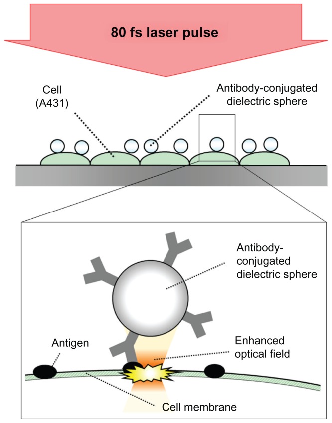
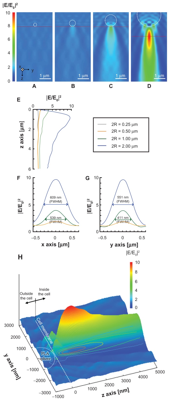
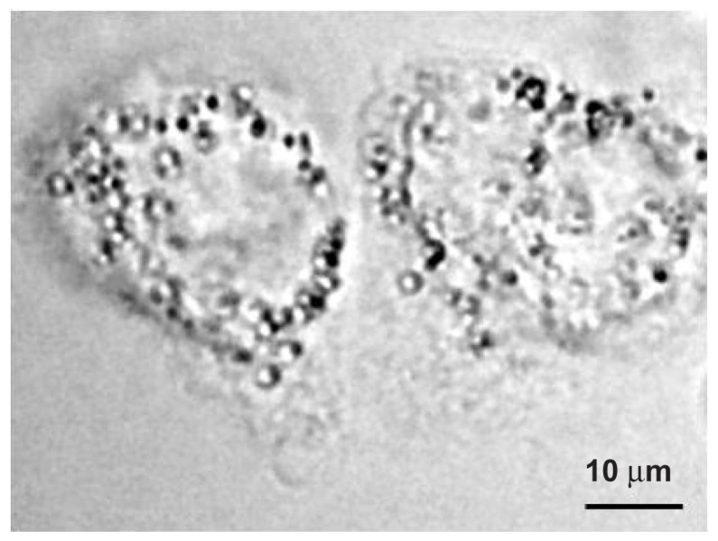
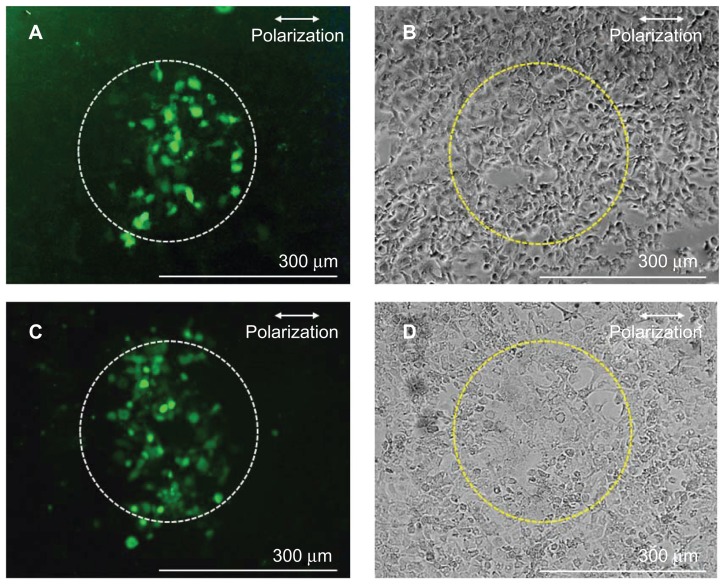
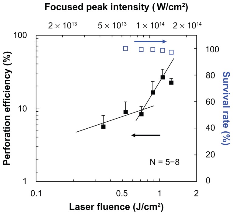
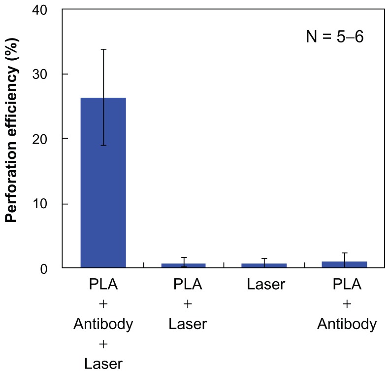
Pulsed laser interaction with small metallic and dielectric particles has been receiving attention as a method of drug delivery to many cells. However, most of the particles are attended by many risks, which are mainly dependent upon particle size. Unlike other widely used particles, biodegradable particles have advantages of being broken down and eliminated by innate metabolic processes. In this paper, the perforation of cell membrane by a focused spot with transparent biodegradable microspheres excited by a single 800 nm, 80 fs laser pulse is demonstrated. A polylactic acid (PLA) sphere, a biodegradable polymer, was used. Fluorescein isothiocyanate (FITC)-dextran and short interfering RNA were delivered into many human epithelial carcinoma cells (A431 cells) by applying a single 80 fs laser pulse in the presence of antibody-conjugated PLA microspheres. The focused intensity was also simulated by the three-dimensional finite-difference time-domain method. Perforation by biodegradable spheres compared with other particles has the potential to be a much safer phototherapy and drug delivery method for patients. The present method can open a new avenue, which is considered an efficient adherent for the selective perforation of cells which express the specific antigen on the cell membrane.

Keywords: biodegradable polymer; drug delivery; femtosecond laser; transfection.


Figures

Figure 1
Figure 1
Conceptual diagram of dielectric sphere-mediated perforation using femtosecond (fs) laser. Notes: Biodegradable spheres are conjugated to cell membrane via antigen– antibody interaction. Femtosecond laser illumination to the spheres generates a strongly enhanced optical field under the sphere for perforation.
Figure 2
Figure 2
(AD) Optical intensity distributions on the yz plane simulated by the three-dimensional finite-difference time-domain method for PLA spheres of different diameters: (A) 250 nm, (B) 500 nm, (C) 1000 nm, and (D) 2000 nm. A plane wave is illuminated to the sphere with the wave vector in the z direction. The incident wave of 800 nm in wavelength is linearly polarized along the x-axis. (EG) Optical intensity distributions along (E) the z-axis under the sphere, (F) the x-axis under the sphere on the peak intensity, shown as red horizontal line in AD, and (G) the y-axis under the sphere on the peak intensity. (H) Relative positions of focused far field, PLA sphere, and cell membrane on yz plane in the case of 2000 nm PLA sphere. Note: Dashed circle and gray plane indicate the positions of the PLA sphere and the cell membrane, respectively. Abbreviations: FWHM, Width at half maximum; PLA, polylactic acid.
Figure 3
Figure 3
Phase contrast image of the A431 cells before laser illumination. Note: The polylactic acid spheres with a diameter of 2000 nm conjugated to the surface of the cells.
Figure 4
Figure 4
Fluorescence (A and C) and phase contrast (B and D) images of A431 cells perforated by using antibody-conjugated polylactic acid spheres irradiated by a single fs laser pulse at 1.06 J/cm2 in the presence of fluorescein isothiocyanate-dextran (A and B) and Alexa Fluor-labeled small interfering RNA (C and D). Note: Dashed circles (300 μm diameter) indicate the laser irradiated area. Abbreviation: fs, femtosecond.
Figure 5
Figure 5
Dependence of perforation efficiency evaluated by using fluorescein isothiocyanate-dextran (closed squares) and survival rate (open squares) on the laser fluence. Notes: A single shot of 80-fs laser pulse was irradiated. The corresponding peak focused intensity under the polylactic acid sphere which was derived from Figure 2 is also shown on the top horizontal axis. Abbreviation: fs, femtosecond.
Figure 6
Figure 6
Average perforation efficiency under four different conditions. Note: A single 80 fs laser pulse was irradiated at 1.06 J/cm2 (1.29 × 10¹⁴ W/cm2 under the PLA sphere). Abbreviations: fs, femtosecond; PLA, polylactic acid.

References

    1. Oliveira MB, Mano JF. Polymer-based microparticles in tissue engineering and regenerative medicine. Biotech Prog. 2011;27(4):897–912. - PubMed
    1. Li W, Brown PK, Wang LV, Xia Y. Gold nanocages as contrast agents for photoacoustic imaging. Contrast Media Mol Imaging. 2011;6(5):370–377. - PMC - PubMed
    1. Bardhan R, Lal S, Joshi A, Halas NJ. Theranostic nanoshells: from probe design to imaging and treatment of cancer. Acc Chem Res. 2011;44(10):936–946. - PMC - PubMed
    1. Singh M, Chakrapani A, O’Hagan D. Nanoparticles and microparticles as vaccine-delivery systems. Expert Rev Vaccines. 2007;6(5):797–808. - PubMed
    1. Paulo CS, Pires das Neves R, Ferreira LS. Nanoparticles for intracellular-targeted drug delivery. Nanotechnology. 2011;22(49):494002/1–11. - PubMed

Publication types

MeSH terms