

Endoscopic management of a primary duodenal carcinoid tumor
- PMID: 22679400
- PMCID: PMC3364033
- DOI: 10.1159/000337870
Endoscopic management of a primary duodenal carcinoid tumor
Abstract
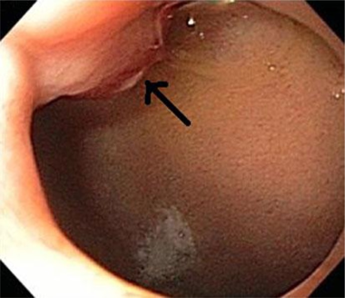
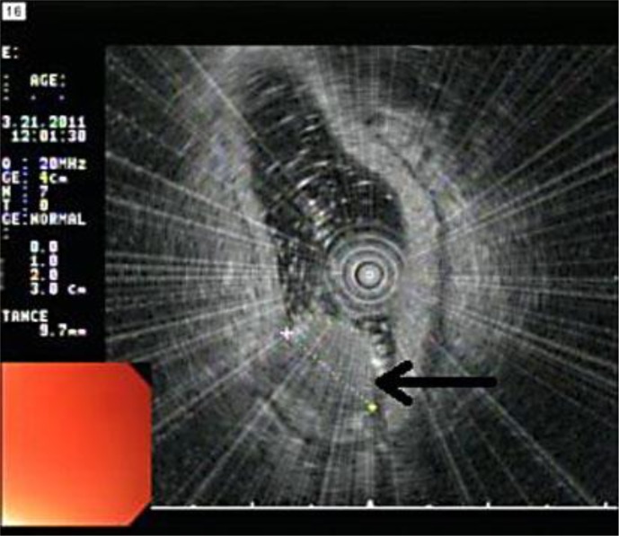
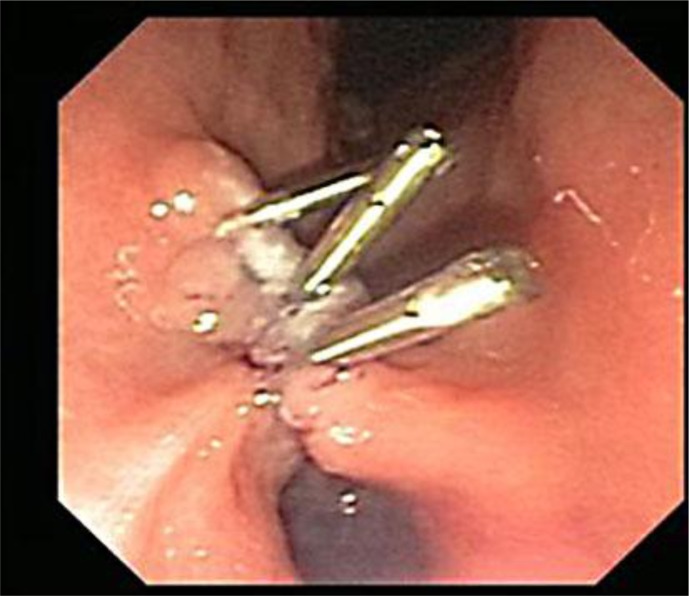
Carcinoids are rare, slow-growing tumors originating from a variety of different neuroendocrine cell types. They are identified histologically by their affinity for silver salts and by positive reactions to neuroendocrine markers such as neuron-specific enolase, synaptophysin and chromogranin. They can present with various clinical symptoms and are difficult to diagnose. We present the case of a 43-year-old woman who was referred for evaluation of anemia. Upper endoscopy showed a duodenal bulb mass around 1 cm in size. Histopathological and immunohistochemistry staining were consistent with the diagnosis of a carcinoid tumor. Further imaging and endoscopic studies showed no other synchronous carcinoid lesions. Endoscopic ultrasound (EUS) revealed a 1 cm lesion confined to the mucosa and no local lymphadenopathy. Successful endoscopic mucosal resection of the mass was performed. Follow-up surveillance 6 months later with EUS and Octreoscan revealed no new lesions suggestive of recurrence. No consensus guidelines exist for the endoscopic management of duodenal carcinoid tumors. However, endoscopic resection is safe and preferred for tumors measuring 1 cm or less with no evidence of invasion of the muscularis layer.
Keywords: Duodenal carcinoid tumors; Endoscopic mucosal resection; Endoscopic ultrasound; Neuroendocrine cell types.
Figures
References
-
- Oberg K. Neuroendocrine gastrointestinal and lung tumors (carcinoid tumors), carcinoid syndrome, and related disorders. In: Melmed S, Polonsky KS, Larsen PR, Kronenberg HM, editors. Williams Textbook of Endocrinology. ed 12. Philadelphia: Elsevier/Saunders; 2011. pp. 1809–1828.
-
- Nikou GC, Toubanakis C, Moulakakis KG, Pavlatos S, Kosmidis C, Mallas E, Safioleas P, Sakorafas GH, Safioleas MC. Carcinoid tumors of the duodenum and ampulla of Vater: current diagnostic and therapeutic approach in a series of 8 patients. Case series. Int J Surg. 2011;9:248–253. - PubMed
-
- Oberg K. Gastrointestinal carcinoid tumors (gastrointestinal neuroendocrine tumors) and the carcinoid syndrome. In: Feldman M, Friedman L, Brandt L, editors. Sleisenger and Fordtran's Gastrointestinal and Liver Disease. ed 9. Philadelphia: Elsevier/Saunders; 2010. pp. 475–490.
-
- World Health Organization . Histological Typing of Endocrine Tumors. Geneva: WHO; 2000. International Classification of Tumors.
Publication types
LinkOut - more resources
Full Text Sources
