. 2012 Jun 5:3:192.
doi: 10.3389/fmicb.2012.00192. eCollection 2012.

Heteroduplex formation, mismatch resolution, and genetic sectoring during homologous recombination in the hyperthermophilic archaeon sulfolobus acidocaldarius

Affiliations

Heteroduplex formation, mismatch resolution, and genetic sectoring during homologous recombination in the hyperthermophilic archaeon sulfolobus acidocaldarius

Dominic Mao et al. Front Microbiol. .

Abstract

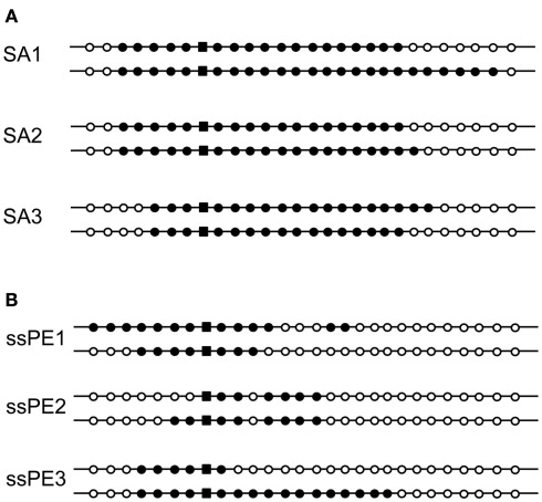
Hyperthermophilic archaea exhibit certain molecular-genetic features not seen in bacteria or eukaryotes, and their systems of homologous recombination (HR) remain largely unexplored in vivo. We transformed a Sulfolobus acidocaldariuspyrE mutant with short DNAs that contained multiple non-selected genetic markers within the pyrE gene. From 20 to 40% of the resulting colonies were found to contain two Pyr(+) clones with distinct sets of the non-selected markers. The dual-genotype colonies could not be attributed to multiple DNAs entering the cells, or to conjugation between transformed and non-transformed cells. These colonies thus appear to represent genetic sectoring in which regions of heteroduplex DNA formed and then segregated after partial resolution of inter-strand differences. Surprisingly, sectoring was also frequent in cells transformed with single-stranded DNAs. Oligonucleotides produced more sectored transformants when electroporated as single strands than as a duplex, although all forms of donor DNA (positive-strand, negative-strand, and duplex) produced a diversity of genotypes, despite the limited number of markers. The marker patterns in the recombinants indicate that S. acidocaldarius resolves individual mismatches through un-coordinated short-patch excision followed by re-filling of the resulting gap. The conversion events that occur during transformation by single-stranded DNA do not show the strand bias necessary for a system that corrects replication errors effectively; similar events also occur in pre-formed heteroduplex electroporated into the cells. Although numerous mechanistic details remain obscure, the results demonstrate that the HR system of S. acidocaldarius can generate remarkable genetic diversity from short intervals of moderately diverged DNAs.

Keywords: gene conversion; genetic transformation; linear DNA; mismatch repair.


Figures

Figure 1
Figure 1
Routes of genetic transformation by non-reciprocal events. All schemes begin with a region of heteroduplex that has formed between a donor (input) DNA and the recipient genome. The donor DNA bears a selectable marker (bar in center) and multiple non-selected markers (dark semi-circles). (A) Incorporation of the donor strand by trimming, gap-filling, and ligation leads to one transformed cell and a non-transformed daughter cell which is lost. (B) Similar incorporation of the donor strand is followed by a large gap on the opposite strand and re-filling, thereby copying all donor markers to the opposite strand (complete conversion) yielding two identical daughter cells. (C) A smaller gap opposite the donor strand results in partial conversion and two distinct daughter cells (genetic sectoring).
Figure 2
Figure 2
Dual-genotype transformants. Diagrams show the marker (donor vs. recipient allele) at each of 28 sites distinguishing the input (donor) pyrE sequence from the chromosomal gene (marker positions are not drawn to scale). For details on the marked sites, see Grogan and Rockwood (2010). The donor allele is represented by a filled dot, the recipient allele is an open circle, and the selected marker is represented by a filled square. Each pair of genotypes was recovered from a colony of cells on selective medium that were transformed to the Pyr+ phenotype by double-stranded pyrEv3 DNA (see Materials and Methods).
Figure 3
Figure 3
Effects of blocking conjugation or transforming with single-stranded DNA. The genotypes of dual-genotype transformants are shown as for Figure 2. (A) Strain SA1 (conjugation-deficient) transformed with the double-stranded pyrEv3 cassette. (B) Strain MR31 transformed with single-stranded pyrEv3 DNA (see Materials and Methods).
Figure 4
Figure 4
Transformation by differentially marked oligonucleotides. Two synthetic DNAs (Table 2), representing the top strand (T) and bottom strand (B) of base pairs 43–227 of pyrE, were electroporated individually (A,B) or as a duplex (C,D). Markers are given in Table 2; the transformants were selected and analyzed as for Figure 2. (C) Shows pairs of genotypes from sectored colonies, whereas (D) shows the single genotypes of non-sectored colonies recovered in the same experiment. Symbols: top-strand donor allele, black; bottom-strand donor allele, gray; recipient allele, white.
Figure 5
Figure 5
Common genotypes formed by differentially marked oligonucleotides. The two or three most frequent genotypes (data of Figure 4) are indicated with their frequency among all recombinants. It should be noted that the number of potential genotypes is much higher for the duplex than for the individual strands of donor DNA, resulting in lower absolute frequencies of the most frequent genotypes. Symbols follow the scheme of Figure 4.
Figure 6
Figure 6
Frequency of strand-specific donor marker incorporation. Graphs show the proportion of recombinants retaining the indicated donor marker at the corresponding position in the synthetic pyrE DNA described in Table 2. Panels and symbols correspond to Figure 4. (A) Sectored transformants from ssDNAs; (B) sectored transformants from dsDNA; (C) non-sectored transformants from dsDNA.
Figure 7
Figure 7
Alternative models ofends-out” recombination. Essential features of two alternatives proposed to explain integration of a linear DNA sequence into a cellular genome by HR. (A) “Double-crossover” (strand exchange) model (see Langston and Symington, 2004); (B) “strand-assimilation” model (see Leung et al., 1997); (C) histogram showing the distribution of the net excess of T over B markers among sectored and non-sectored (simple) recombinants from the differentially marked dsDNA.
Figure 8
Figure 8
Mechanistic alternatives for “ends-out” HR in Sulfolobus. (A) ssDNA annealing at a transient gap in the chromosome; (B) ssDNA annealing to a transiently regressed replication fork; (C) dsDNA incorporated at a gap in the chromosome, or alternatively, at a regressed replication fork (dotted arrow). Arrowheads indicate 3 ends, black bars correspond to the top (sense) strand of the pyrE gene, gray bars indicate the bottom (anti-sense) strand.

Similar articles

Cited by

References

    1. Ajon M., Frols S., Van Wolferen M., Stoecker K., Teichmann D., Driessen A. J., Grogan D. W., Albers S. V., Schleper C. (2011). UV-inducible DNA exchange in hyperthermophilic archaea mediated by type IV pili. Mol. Microbiol. 82, 807–81710.1111/j.1365-2958.2011.07861.x - DOI - PubMed
    1. Aylon Y., Kupiec M. (2004). DSB repair: the yeast paradigm. DNA Repair (Amst.) 3, 797–81510.1016/j.dnarep.2004.04.013 - DOI - PubMed
    1. Barnes R. L., McCulloch R. (2007). Trypanosoma brucei homologous recombination is dependent on substrate length and homology, though displays a differential dependence on mismatch repair as substrate length decreases. Nucleic Acids Res. 35, 3478–349310.1093/nar/gkm249 - DOI - PMC - PubMed
    1. Beattie T. R., Bell S. D. (2012). Coordination of multiple enzyme activities by a single PCNA in archaeal Okazaki fragment maturation. EMBO J. 31, 1556–156710.1038/emboj.2012.12 - DOI - PMC - PubMed
    1. Bernander R., Poplawski A. (1997). Cell cycle characteristics of thermophilic archaea. J. Bacteriol. 179, 4963–4969 - PMC - PubMed