

Genetics of host response to Leishmania tropica in mice - different control of skin pathology, chemokine reaction, and invasion into spleen and liver
- PMID: 22679519
- PMCID: PMC3367980
- DOI: 10.1371/journal.pntd.0001667
Genetics of host response to Leishmania tropica in mice - different control of skin pathology, chemokine reaction, and invasion into spleen and liver
Abstract
Background: Leishmaniasis is a disease caused by protozoan parasites of genus Leishmania. The frequent involvement of Leishmania tropica in human leishmaniasis has been recognized only recently. Similarly as L. major, L. tropica causes cutaneous leishmaniasis in humans, but can also visceralize and cause systemic illness. The relationship between the host genotype and disease manifestations is poorly understood because there were no suitable animal models.
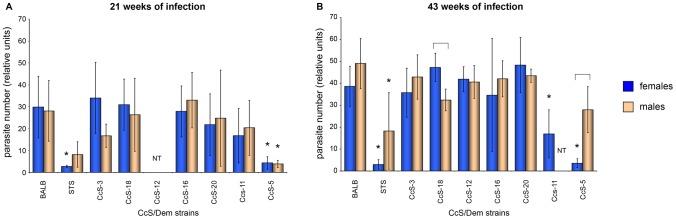
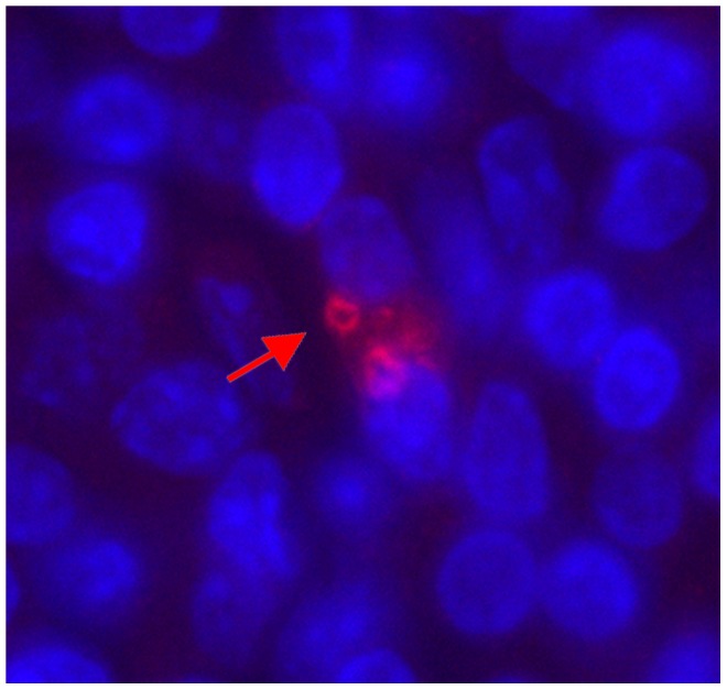
Methods: We studied susceptibility to L. tropica, using BALB/c-c-STS/A (CcS/Dem) recombinant congenic (RC) strains, which differ greatly in susceptibility to L. major. Mice were infected with L. tropica and skin lesions, cytokine and chemokine levels in serum, and parasite numbers in organs were measured.
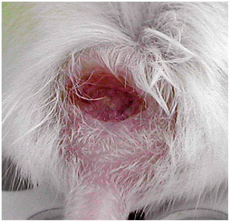
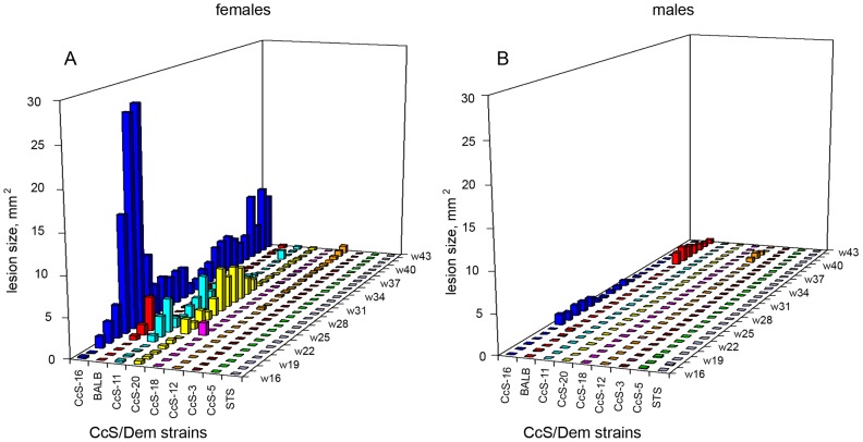
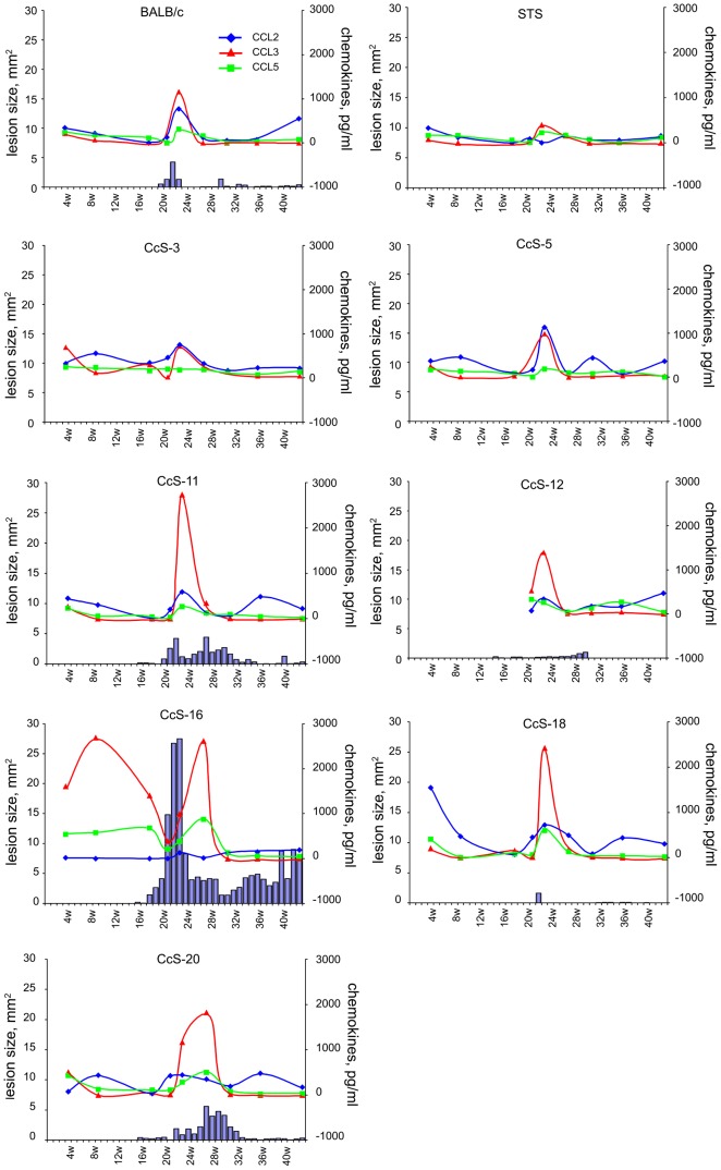
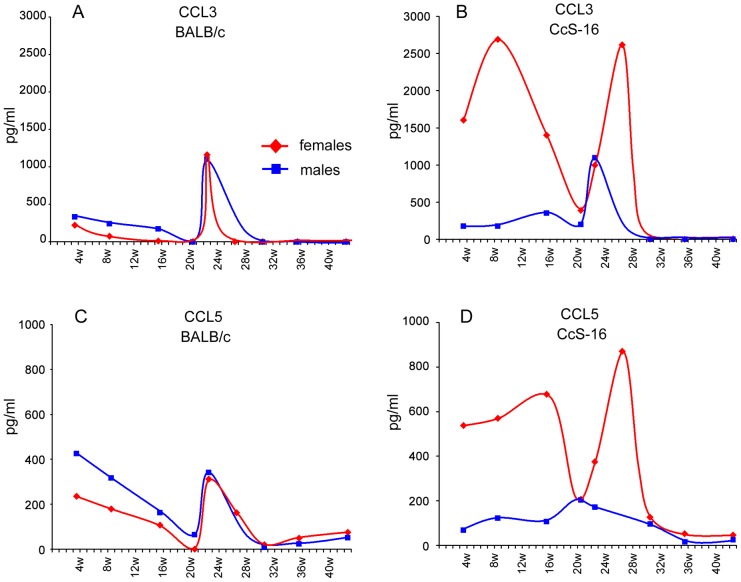
Principal findings: Females of BALB/c and several RC strains developed skin lesions. In some strains parasites visceralized and were detected in spleen and liver. Importantly, the strain distribution pattern of symptoms caused by L. tropica was different from that observed after L. major infection. Moreover, sex differently influenced infection with L. tropica and L. major. L. major-infected males exhibited either higher or similar skin pathology as females, whereas L. tropica-infected females were more susceptible than males. The majority of L. tropica-infected strains exhibited increased levels of chemokines CCL2, CCL3 and CCL5. CcS-16 females, which developed the largest lesions, exhibited a unique systemic chemokine reaction, characterized by additional transient early peaks of CCL3 and CCL5, which were not present in CcS-16 males nor in any other strain.
Conclusion: Comparison of L. tropica and L. major infections indicates that the strain patterns of response are species-specific, with different sex effects and largely different host susceptibility genes.
Conflict of interest statement
The authors have declared that no competing interests exist.
Figures
Similar articles
-
Leishmania tropica: suggestive evidences for the effect of infectious dose on pathogenicity and immunogenicity in an experimental model.Parasitol Res. 2018 Sep;117(9):2949-2956. doi: 10.1007/s00436-018-5991-7. Epub 2018 Jul 5. Parasitol Res. 2018. PMID: 29978420
-
Mapping the genes for susceptibility and response to Leishmania tropica in mouse.PLoS Negl Trop Dis. 2013 Jul 11;7(7):e2282. doi: 10.1371/journal.pntd.0002282. Print 2013. PLoS Negl Trop Dis. 2013. PMID: 23875032 Free PMC article.
-
Heterogeneity of humoral immune response to Leishmania tropica in an experimental model.Parasitol Res. 2019 Apr;118(4):1231-1237. doi: 10.1007/s00436-019-06256-3. Epub 2019 Feb 19. Parasitol Res. 2019. PMID: 30778754
-
Leishmania tropica: What we know from its experimental models.Adv Parasitol. 2019;104:1-38. doi: 10.1016/bs.apar.2018.11.001. Epub 2018 Dec 23. Adv Parasitol. 2019. PMID: 31030767 Review.
-
Role of host genetics and cytokines in Leishmania infection.Cytokine. 2021 Nov;147:155244. doi: 10.1016/j.cyto.2020.155244. Epub 2020 Oct 12. Cytokine. 2021. PMID: 33059974 Review.
Cited by
-
Gene-Specific Sex Effects on Susceptibility to Infectious Diseases.Front Immunol. 2021 Oct 14;12:712688. doi: 10.3389/fimmu.2021.712688. eCollection 2021. Front Immunol. 2021. PMID: 34721380 Free PMC article. Review.
-
Analysis of localized immune responses reveals presence of Th17 and Treg cells in cutaneous leishmaniasis due to Leishmania tropica.BMC Immunol. 2013 Nov 22;14:52. doi: 10.1186/1471-2172-14-52. BMC Immunol. 2013. PMID: 24267152 Free PMC article.
-
Deception and manipulation: the arms of leishmania, a successful parasite.Front Immunol. 2014 Oct 20;5:480. doi: 10.3389/fimmu.2014.00480. eCollection 2014. Front Immunol. 2014. PMID: 25368612 Free PMC article. Review.
-
Genotype-driven sensitivity of mice to tick-borne encephalitis virus correlates with differential host responses in peripheral macrophages and brain.J Neuroinflammation. 2025 Jan 28;22(1):22. doi: 10.1186/s12974-025-03354-1. J Neuroinflammation. 2025. PMID: 39875898 Free PMC article.
-
Visualization of Leishmania tropica Infection in BALB/c Mice by Bioluminescence Imaging.Iran Biomed J. 2020 May;24(3):164-72. doi: 10.29252/ibj.24.3.164. Epub 2019 Dec 1. Iran Biomed J. 2020. PMID: 31952434 Free PMC article.
References
-
- Rittig MG, Bogdan C. Leishmania-host-cell interaction: complexities and alternative views. Parasitol Today. 2000;16:292–297. - PubMed
-
- Reiner SL, Locksley RM. The regulation of immunity to Leishmania major. Annu Rev Immunol. 1995;13:151–177. - PubMed
-
- McMahon-Pratt D, Alexander J. Does the Leishmania major paradigm of pathogenesis and protection hold for New World cutaneous leishmaniases or the visceral disease? Immunol Rev. 2004;201:206–224. - PubMed
-
- Farrell JP. Leishmania. Boston, Dordrecht, London: Kluwer Academic Publishers; 2002. 193
-
- Kobets T, Grekov I, Lipoldová M. Leishmaniasis: prevention, parasite detection and treatment. Curr Med Chem. 2012;19:1443–1474. - PubMed
Publication types
MeSH terms
Substances
LinkOut - more resources
Full Text Sources
