Fluoroscope guided epidural needle insertioin in midthoracic region: clinical evaluation of Nagaro's method
- PMID: 22679541
- PMCID: PMC3366311
- DOI: 10.4097/kjae.2012.62.5.441
Fluoroscope guided epidural needle insertioin in midthoracic region: clinical evaluation of Nagaro's method
Abstract
Background: In the midthoracic region, a fluroscope guided epidural block has been proposed by using a pedicle as a landmark to show the height of the interlaminar space (Nagaro's method). However, clinical implication of this method was not fully evaluated. We studied the clinical usefulness of a fluoroscope guided thoracic epidural block in the midthoracic region.
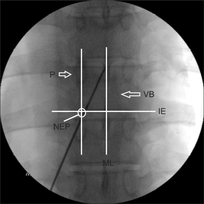
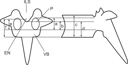
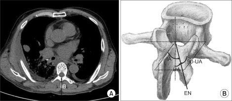
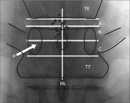
Methods: Twenty four patients were scheduled to receive an epidural block at the T6-7 intervertebral space. The patients were placed in the prone position. The needle entry point was located at the junction between midline of the pedicle paralleled to the midline of the T7 vertebral body (VB) and the lower border of T7 VB on anteroposterior view of the fluoroscope. The needle touched and walked up the lamina, and the interlaminar space (ILS) was sought near the midline of the VB at the height of the pedicle.
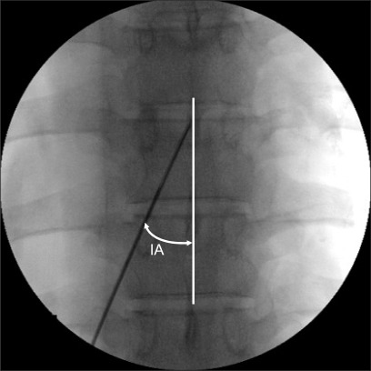
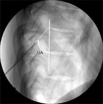
Results: The authors could not insert an epidural needle at T6-7 ILS in two patients and it was instead inserted at T5-6 ILS. However, other patients showed easy insertion at T6-7 ILS. The mean inward and upward angulations were 25° and 55° respectively. The mean actual depth and calculated depth from skin to thoracic epidural space were 5.1 cm and 6.1 cm respectively. Significant correlation between actual needle depth and body weight, podendal index (kg/m) or calculated needle depth was noted.
Conclusions: The fluorposcope guided epidural block by Nagaro's method was useful in the midthoracic region. However, further study for the caudal shift of needle entry point may be needed.
Keywords: Analgesia; Epidural; Fluoroscopy; Thoracic vertebrae.
Figures
References
-
- De Cosmo G, Aceto P, Gualtieri E, Congedo E. Analgesia in thoracic surgery: review. Minerva Anestesiol. 2009;75:393–400. - PubMed
-
- Sawchuk CW, Ong B, Unruh HW, Horan TA, Greengrass R. Thoracic versus lumbar epidural fentanyl for postthoracotomy pain. Ann Thorac Surg. 1993;55:1472–1476. - PubMed
-
- Lomessy A, Magnin C, Viale JP, Motin J, Cohen R. Clinical advantages of fentanyl given epidurally for postoperative analgesia. Anesthesiology. 1984;61:466–469. - PubMed
-
- Hood DD, Dewan DM. Anesthetic and obstetric outcome in morbidly obese parturients. Anesthesiology. 1993;79:1210–1218. - PubMed
-
- Konrad C, Schüpfer G, Wietlisbach M, Gerber H. Learning manual skills in anesthesiology: Is there a recommended number of cases for anesthetic procedures? Anesth Analg. 1998;86:635–639. - PubMed
LinkOut - more resources
Full Text Sources