. 2012 May;62(5):461-7.
doi: 10.4097/kjae.2012.62.5.461. Epub 2012 May 24.

The effects of hydrogen sulfide under sevoflurane administration against ischemia and reperfusion injury in isolated rat heart

Affiliations

The effects of hydrogen sulfide under sevoflurane administration against ischemia and reperfusion injury in isolated rat heart

Dong Kyu Lee et al. Korean J Anesthesiol. 2012 May.

Abstract

Background: Hydrogen sulfide (H(2)S) produces a protective effect against myocardial ischemia and reperfusion injury. Sevoflurane, which is used for anesthesia in cardiac problem patients, also has a protective effect. This study is designed to reveal the effects of H(2)S under sevoflurane using rat hearts.

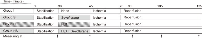
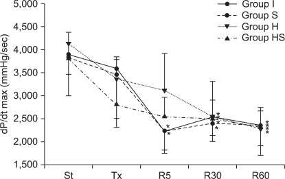
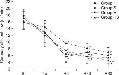
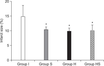
Methods: The hearts were Langendorff-perfused, subjected to 30 minutes ischemia and 60 minutes reperfusion. Group I was a control group. The other groups were pretreated for 15 minutes before ischemia as follows: 1.6% sevoflurane for group S; 18.5 µM H(2)S S for group H; and 1.6% sevoflurane and 18.5 µM H(2)S simultaneously for group HS. Hemodynamics and the infarct size were measured.

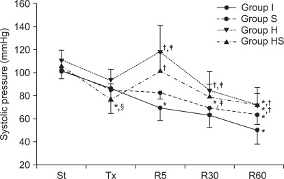
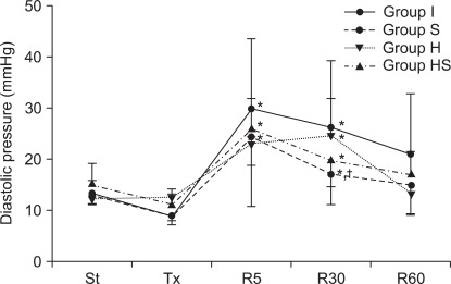
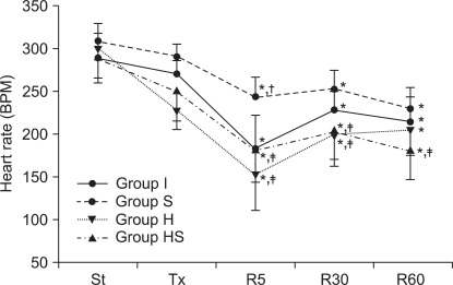
Results: Group HS presented depressed hemodynamics during pretreatment. LV function in group HS achieved better recovery than group I after reperfusion. The infarct size of groups S, H and HS was smaller than group I, while there were no differences between groups S, H and HS.

Conclusions: Exogenous H(2)S did not enhance the preconditioning effects of sevoflurane. Rather, the results suggest that H(2)S under sevoflurane might depress hemodynamics.

Keywords: Heart; Hydrogen sulfide; In vitro; Ischemia; Reperfusion injury; Sevoflurane.


Figures

Fig. 1
Fig. 1
Experimental protocol. After stabilization, each group was subjected to 30 minutes of global ischemia and 60 minutes of reperfusion. Before global ischemia, a 15 minute pre-treatment period was conducted and specific agents were applied to each group as follows: perfusate and mixed gas (95% O2 + 5% CO2) only for group I; mixed gas with 1.6% sevoflurane and perfusate without H2S for group S; 18.5 µM H2S mixed perfusate and mixed gas for group H; 1.6% sevoflurane and 18.5 µM H2S for group HS. Hemodyamic variables were measured after stabilization, after pre-treatment and 5, 30, 60 minutes after reperfusion started (Arrows). After all processes were completed, triphenyl tetrazolium staining was performed to calculate infarct size.
Fig. 2
Fig. 2
Left ventricular end systolic pressure. Group HS showed lower systolic pressure after pre-treatment than those after stabilization. Groups H and HS showed mostly higher pressures during the reperfusion period than groups I and S. St: after stabilization, Tx: pretreatment, R5: reperfusion 5 minutes, R30: reperfusion 30 minutes, R60: reperfusion 60 minutes. P < 0.05 is remarked as follows: *vs. after stabilization in the same group. vs. Group I, vs. Group S, §vs. Group H at the same measurement time.
Fig. 3
Fig. 3
Left ventricular diastolic pressure. During the reperfusion period, diastolic pressure in all groups initially increased, and then progressively decreased over time. In group H, diastolic pressure was sustained at a high rate until 30 minutes after reperfusion and then decreased toward the value of that after stabilization. Groups S and HS showed similar patterns in pressure changes. St: after stabilization, Tx: pretreatment, R5: reperfusion 5 minutes, R30: reperfusion 30 minutes, R60: reperfusion 60 minutes. P < 0.05 is remarked as follows: *vs. after stabilization and/or pretreatment in the same group, vs. groups I and H at the same measurement time.
Fig. 4
Fig. 4
Heart rate. Heart rates during the reperfusion period in all groups were lower than those prior to ischemia. Heart rates in groups H and HS were mostly lower than those in group S during the reperfusion period. St: after stabilization, Tx : pretreatment, R5: reperfusion 5 minutes, R30: reperfusion 30 minutes, R60: reperfusion 60 minutes. P < 0.05 is remarked as follows: *vs. after stabilization in the same group, vs. groups I, vs. group S at the same measurement time.
Fig. 5
Fig. 5
Maximum rate of change of left ventricular pressure (dP/dt max). 5 minutes after reperfusion, dP/dt max in groups I and S decreased rapidly. During the rest of reperfusion, dP/dt max of all groups remained low. St: after stabilization, Tx: pretreatment, R5: reperfusion 5 minutes, R30: reperfusion 30 minutes, R60: reperfusion 60 minutes. P < 0.05 is remarked as follows: *vs. after stabilization in the same group.
Fig. 6
Fig. 6
Coronoary effluent flows measured in rate. During the reperfusion period, coronary effluent flow rates of all groups decreased with time. Primarily, group S showed higher rates than groups I, H and HS. St: after stabilization, Tx: pretreatment, R5: reperfusion 5 minutes, R30: reperfusion 30 minutes, R60: reperfusion 60 minutes. P < 0.05 is remarked as follows: *vs. after stabilization in the same group. At the same measuring time, vs. groups I, vs. group S at the same measurement time.
Fig. 7
Fig. 7
Infarct size measured in each group. Groups S, H and HS showed smaller infarct size than group I. There was no difference between infarct size of groups S, H and HS. P < 0.05 is remarked as follows: *vs. group I.

Similar articles

References

    1. Vaage J, Jensen U, Ericsson A. Neurologic injury in cardiac surgery: aortic atherosclerosis emerges as the single most important risk factor. Scand Cardiovasc J. 2000;34:550–557. - PubMed
    1. Valen G, Vaage J. Pre- and postconditioning during cardiac surgery. Basic Res Cardiol. 2005;100:179–186. - PubMed
    1. Venugopal V, Ludman A, Yellon DM, Hausenloy DJ. 'Conditioning' the heart during surgery. Eur J Cardiothorac Surg. 2009;35:977–987. - PubMed
    1. Weber NC, Schlack W. Inhalational anaesthetics and cardioprotection. Handb Exp Pharmacol. 2008;182:187–207. - PubMed
    1. Wang R. Two's company, three's a crowd: can H2S be the third endogenous gaseous transmitter? FASEB J. 2002;16:1792–1798. - PubMed

LinkOut - more resources