

Molecular techniques for pathogen identification and fungus detection in the environment
- PMID: 22679603
- PMCID: PMC3359816
- DOI: 10.5598/imafungus.2011.02.02.09
Molecular techniques for pathogen identification and fungus detection in the environment
Abstract
Many species of fungi can cause disease in plants, animals and humans. Accurate and robust detection and quantification of fungi is essential for diagnosis, modeling and surveillance. Also direct detection of fungi enables a deeper understanding of natural microbial communities, particularly as a great many fungi are difficult or impossible to cultivate. In the last decade, effective amplification platforms, probe development and various quantitative PCR technologies have revolutionized research on fungal detection and identification. Examples of the latest technology in fungal detection and differentiation are discussed here.
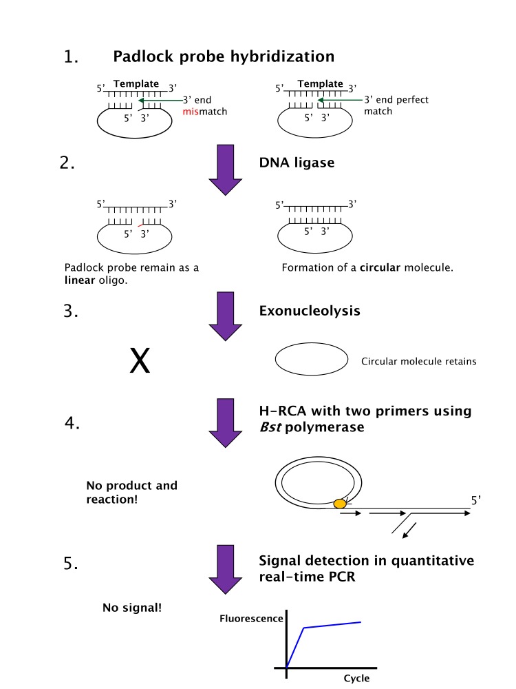
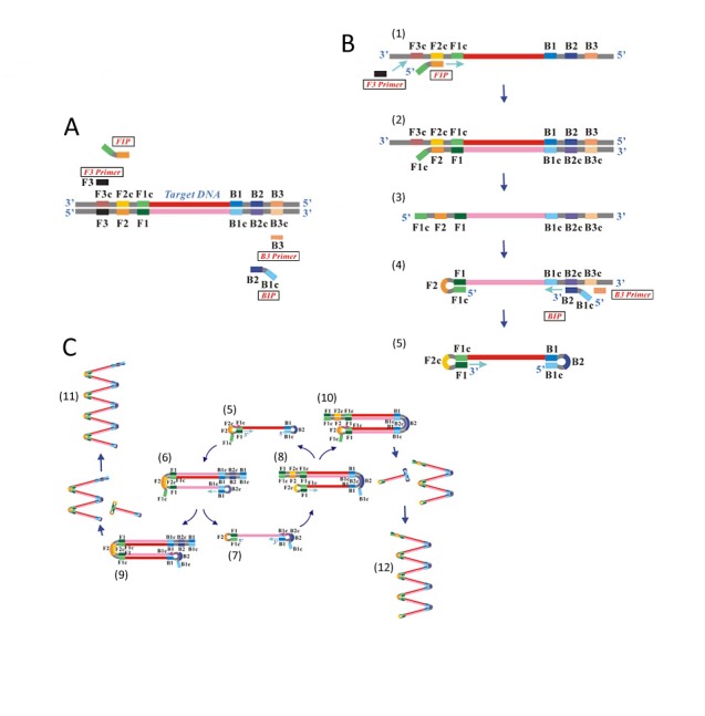
Keywords: FISH; LAMP; macroarray; medical mycology; molecular diagnostics; molecular ecology; padlock probe; pathogenic fungi; plant pathology; rolling circle amplification.
Figures
Similar articles
-
Multiplex and quantifiable detection of nucleic acid from pathogenic fungi using padlock probes, generic real time PCR and specific suspension array readout.J Microbiol Methods. 2009 Aug;78(2):195-202. doi: 10.1016/j.mimet.2009.05.016. Epub 2009 May 30. J Microbiol Methods. 2009. PMID: 19490930
-
Isothermal nucleic acid amplification techniques for detection and identification of pathogenic fungi: A review.Mycoses. 2020 Oct;63(10):1006-1020. doi: 10.1111/myc.13140. Epub 2020 Aug 23. Mycoses. 2020. PMID: 32648947 Review.
-
Fungal molecular diagnostics: a mini review.J Appl Genet. 2004;45(1):3-15. J Appl Genet. 2004. PMID: 14960763 Review.
-
High specific and ultrasensitive isothermal detection of microRNA by padlock probe-based exponential rolling circle amplification.Anal Chem. 2013 Aug 20;85(16):7941-7. doi: 10.1021/ac401715k. Epub 2013 Jul 31. Anal Chem. 2013. PMID: 23855808
-
Recent Advances in Molecular Diagnostics of Fungal Plant Pathogens: A Mini Review.Front Cell Infect Microbiol. 2021 Jan 11;10:600234. doi: 10.3389/fcimb.2020.600234. eCollection 2020. Front Cell Infect Microbiol. 2021. PMID: 33505921 Free PMC article. Review.
Cited by
-
A Simple and Rapid Fungal DNA Isolation Assay Based on ZnO Nanoparticles for the Diagnosis of Invasive Aspergillosis.Micromachines (Basel). 2020 May 19;11(5):515. doi: 10.3390/mi11050515. Micromachines (Basel). 2020. PMID: 32438738 Free PMC article.
-
Current ecological understanding of fungal-like pathogens of fish: what lies beneath?Front Microbiol. 2014 Feb 19;5:62. doi: 10.3389/fmicb.2014.00062. eCollection 2014. Front Microbiol. 2014. PMID: 24600442 Free PMC article. Review.
-
The PHD transcription factor Cti6 is involved in the fungal colonization and aflatoxin B1 biological synthesis of Aspergillus flavus.IMA Fungus. 2021 May 18;12(1):12. doi: 10.1186/s43008-021-00062-2. IMA Fungus. 2021. PMID: 34006318 Free PMC article.
-
Development of a Novel Diagnostic Tool for Cercospora Species Based on BOX-PCR System.Curr Microbiol. 2022 Aug 16;79(10):290. doi: 10.1007/s00284-022-02989-0. Curr Microbiol. 2022. PMID: 35972567
-
Emerging Fungal Pathogens Warfare, Fungi-Like Organisms, and the World's Major Crops: Lessons from the Past and Solutions for the Future.Curr Microbiol. 2025 Aug 6;82(9):440. doi: 10.1007/s00284-025-04406-8. Curr Microbiol. 2025. PMID: 40768075 Review.
References
-
- Agindotan B, Perry KL. (2007) Macroarray detection of plant RNA viruses using randomly primed and amplified complementary DNAs from infected plants. Phytopathology 97: 119–127 - PubMed
-
- Agindotan B, Perry KL. (2008) Macroarray detection of eleven potato-infecting viruses and Potato spindle tuber viroid. Plant Disease 92: 730–740 - PubMed
-
- Agrios G. (2005) Plant Pathology. 5th edn London: Elsevier Academic Press;
LinkOut - more resources
Full Text Sources
Other Literature Sources