Review
. 2012;19(22):3748-62.
doi: 10.2174/092986712801661130.

Homeostasis and the importance for a balance between AKT/mTOR activity and intracellular signaling

Affiliations
Review

Homeostasis and the importance for a balance between AKT/mTOR activity and intracellular signaling

D A Altomare et al. Curr Med Chem. 2012.

Abstract

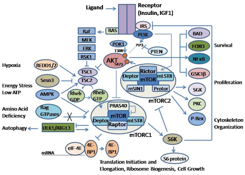
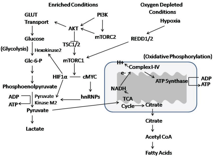
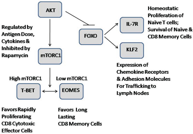
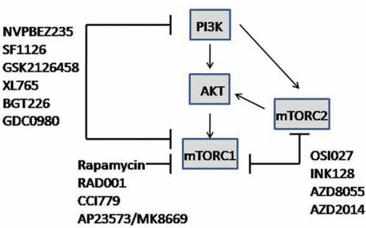
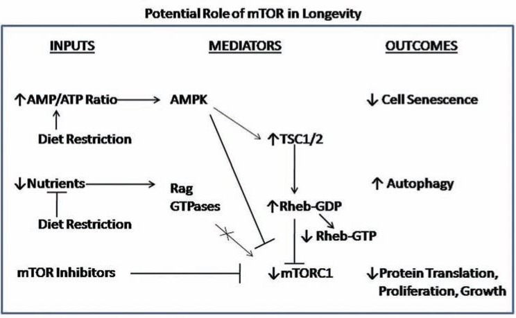
The AKT family of serine threonine kinases is of critical importance with regard to growth factor signaling, cell proliferation, survival and oncogenesis. Engagement of signaling receptors induces the lipid kinase, phosphatidylinositol 3-kinase (PI3K), which enables the activation of AKT. Responsive to the PI3K/AKT pathway is the mammalian target of rapamycin (mTOR), a major effector that is specifically implicated in the regulation of cell growth as a result of nutrient availability and cellular bioenergetics. These kinases mediate the activity of a multitude of intracellular signaling molecules and intersect with multiple pathways that regulate cellular processes. Elucidating the role of AKT/mTOR in metabolism and in hallmark signaling pathways that are aberrantly affected in cancer has provided a solid foundation of discoveries. From this, new research directions are emerging with regard to the role of AKT/mTOR in diabetes and T cell-mediated immunity. As a result, a new perspective is developing in how AKT/mTOR functions within intracellular signaling pathways to maintain cellular homeostasis. An appreciation is emerging that altered equilibrium of AKT/mTOR pathways contributes to disease and malignancy. Such new insights may lead to novel intervention strategies that may be useful to reprogram or reset the balance of intracellular signaling.


Figures

Fig. (1)
Fig. (1)
Schematic diagram of the AKT/mTOR signaling pathway. Insulin or insulin growth factor-1 (IGF-1) stimulation is shown to represent growth factor signaling. Upon activation the insulin receptor (IR) phosphorylates insulin receptor substrate (IRS), which activates phosphatidylinositol 3-kinase (PI3K), which in turns phosphorylates phosphoinositides to generate phosphatidylinositol (3,4,5)-trisphosphate (PIP3). Phosphatase and tensin homolog (PTEN) can dephosphorylate PIP3 to regulate activity of the signaling pathway. AKT is activated by binding PIP3 to it the amino terminal pleckstrin homology (PH) domain, which promotes translocation of AKT to the plasma membrane (represented by a dotted line) where the carboxyl terminal Thr308 is phosphorylated by phosphoinositide-dependent kinase-1 (PDK1), and Ser473 is phosphorylated by mTORC2. AKT regulates several cellular processes such as survival and cell proliferation through a variety of downstream proteins, including BAD (Bcl-2-antagonist of cell death), FOXO (Forkhead Box O), NFćB, and GSK-3β (glycogen synthase kinase 3-beta), among others. AKT can directly phosphorylate and inactivate a 40kDa, proline-rich protein (PRAS40), to relieve the inhibitory regulation on mTORC1 activity. AKT also can phosphorylate and inactivate the tuberous sclerosis (TSC) tumor suppressor protein complex, which acts as a GTPase-activating protein for the RAS-related small G protein (Rheb) to regulate its activity. Retention of the Rheb-GTP form activates mTOR. mTOR is complexed with other proteins including Raptor, mLST8 and Deptor in complex I (mTORC1) and Rictor, mLST8, Deptor, Sin1 and Protor in complex II (mTORC2). mTORC1 regulation is mediated through a variety of environmental signals, which are mediated by proteins such as REDD (regulated in development and DNA damage responses 1), Sestrin 3 (Sesn3), AMP-activated protein kinase (AMPK) and Rag GTPases. mTORC1 phosphorylates downstream p70S6 kinase (S6K) and the eukaryotic initiation factor 4E-binding protein (4E-BP1), which releases it from inhibiting eIF4E so that 40S ribosomal subunit can be recruited to the 5’ end of mRNAs to initiate protein translation. S6K phosphorylates ribosomal protein S6, which is also involved in regulating protein translation through the 40S ribosomal subunit. mTORC1 is also instrumental in regulating autophagy through ULK1 and ATG13 (autophagy related 13 homolog). In contrast, mTORC2 regulation is still being investigated, although it is known to be activated by growth factor signaling pathways. mTORC2 phosphorylates a different set of proteins, including serum- and glucocorticoid-inducible kinase (SGK), protein kinase C (PKC) and the guanine nucleotide exchange factor Phosphatidylinositol (3,4,5)-Triphosphate-Dependent Rac Exchanger 1 (P-Rex1) to regulate Rac mediated cytoskeleton changes. Importantly, mTORC2 phosphorylates AKT as a feedback into the pathway. Phosphorylation of S6K downstream of mTORC1 is a negative feedback to inhibit IRS signaling and Rictor phosphorylation. The growth factor mediated activation of the RAS/Raf/MEK/ERK/RSK1 pathway is another mechanism whereby there can be crosstalk regulation of the AKT/mTOR signaling pathway.
Fig. (2)
Fig. (2)
AKT/mTOR signaling and crosstalk with glycolysis and mitochondrial oxidative phosphorylation. Glucose is transported into the cells using GLUT transporters. Glycolysis uses glucose to make pyruvate, which can be converted to lactate, or used to generate ATP through oxidative phosphorylation. Oxidative phosphorylation occurs in the mitochondria. The process converts pyruvate to acetyl coenzyme A (acetyl-CoA), and uses the tricarboxylic-acid (TCA) cycle to produce free electrons (e-) that are carried by NADH to the electron-transport chain, resulting in a movement of protons (H+) out of the mitochondrial matrix to establish a electrochemical potential that cumulates in the activation of ATP synthase to synthesize ATP. AKT/mTOR signaling interacts with these metabolic processes through the regulation of GLUT transport. Moreover, hypoxia-inducible factor 1 alpha (HIF1α) is a downstream effector of mTORC1 that is important in upregulating the expression of hexokinase 2 and pyruvate kinase M2, two key rate-limiting enzymes in the glycolytic pathway. Another mechanism downstream of mTORC2 is mediated by cMYC, which activates heterogeneous nuclear ribonucleoproteins (hnRNPs) in the transcription of pyruvate kinase M2.
Fig. (3)
Fig. (3)
Role of AKT/mTOR signaling in the regulation of T cell differentiation, trafficking and survival. mTORC1 regulates the balance of T-bet and Eomes transcription factors, which are known regulators of effector versus memory CD8 T cell function. T-bet is required for the cytotoxic functions and the production of effector cytokines. Eomes expression is required for the memory. Furthermore, inhibition of FOXO1 by AKT phosphorylation results in the upregulation of the expression of Krüppel-like factor 2 (KFL2) to increase CD8 T cell migration and in the expression of interleukin-7 receptor (IL-7R), which is important for the survival of naïve and memory CD8 cells. Figure based on that of Finlay and Cantrell [39].
Fig. (4)
Fig. (4)
General diagram of rapalogs, ATP-competitive mTOR inhibitors and dual AKT/mTOR pathway inhibitors that are clinical use. Primary source was Zhang et al. [80].
Fig. (5)
Fig. (5)
Schematic diagram of how mTOR may play a role in longevity. Calorie or dietary restriction is postulated to increase the AMP/ATP ratio as a result of diminished energy production and decreased nutrient availability. mTOR inhibitors such as rapamycin may be considered mimics for calorie or dietary restriction because ultimately they reduce mTORC1 activity, similar to the affect of AMPK on inhibitory mechanisms to regulate mTORC1, and inhibition by nutrient deprivation of the Rag GTPases, which would normally stimulate mTORC1. As a consequence of diminished mTORC1 activity, protein translation, proliferation and overall growth would be limited and the dynamics of decreased mTORC1 would favor increased autophagy to sustain cell survival and prevent cell senescence.

Similar articles

Cited by

References

    1. Bhaskar P T, Hay N. The two TORCs and Akt. Dev Cell. 2007;12 (4 ): 487–502. - PubMed
    1. Manning B D, Cantley L C. AKT/PKB signaling: navigating downstream. Cell. 2007;129 (7 ):1261–1274. - PMC - PubMed
    1. Franke T F. PI3K/Akt: getting it right matters. Oncogene. 2008;27:6473–6488. - PubMed
    1. Mahajan K, Mahajan N P. PI3K-independent AKT activation in cancers: A treasure trove for novel therapeutics. J Cell Physiol. 2012. Feb 3, [Epub ahead of print] - PMC - PubMed
    1. Mahajan K, Coppola D, Chen Y A, Zhu W, Lawrence H R, Lawrence N J, Mahajan N P. Ack1 Tyrosine Kinase Activation Correlates with Pancreatic Cancer Progression. Am J Pathol. 2012;180 (4 ): 1386–1393. - PMC - PubMed

Publication types

MeSH terms

Substances