

doi: 10.1016/j.str.2012.04.010.
Fusion partner toolchest for the stabilization and crystallization of G protein-coupled receptors
Affiliations
- PMID: 22681902
- PMCID: PMC3375611
- DOI: 10.1016/j.str.2012.04.010
Item in Clipboard
Fusion partner toolchest for the stabilization and crystallization of G protein-coupled receptors
Structure.
.
Abstract
Structural studies of human G protein-coupled receptors (GPCRs) have recently been accelerated through the use of a fusion partner that was inserted into the third intracellular loop. Using chimeras of the human β(2)-adrenergic and human A(2A) adenosine receptors, we present the methodology and data for the initial selection of an expanded set of fusion partners for crystallizing GPCRs. In particular, use of the thermostabilized apocytochrome b(562)RIL as a fusion partner displays certain advantages over previously utilized fusion proteins, resulting in a significant improvement in stability and structure of GPCR-fusion constructs.
Copyright © 2012 Elsevier Ltd. All rights reserved.
Figures
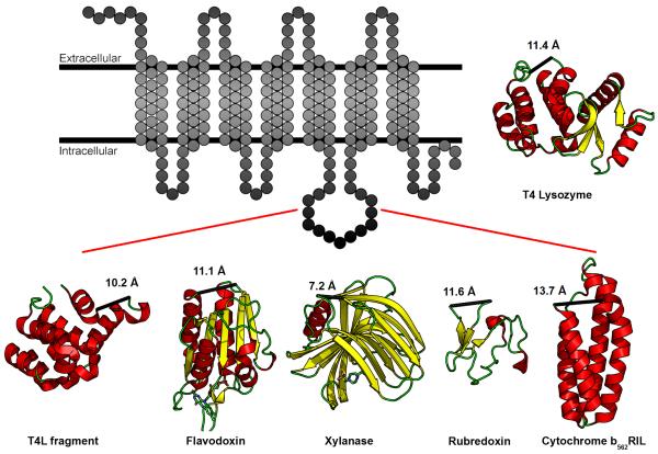
Figure illustrating the insertion of five new domains into the ICL3 of a prototypical GPCR, represented as a transmembrane snakeplot. The five domains are a C-terminal fragment of T4L (PDB ID 2O7A, MW 15.9 kD), flavodoxin (PDB ID 1I1O, MW 14.9 kD), xylanase (PDB ID 2B45, MW 19.1 kD), rubredoxin (PDB ID 1FHM, MW 5.5 kD), and cytochrome b562RIL (PDB ID 1M6T, MW 10.9). These domains exhibit a variety of secondary structures consisting of either α-helices, β-sheets, or a combination of both. Numbers indicate distance (Å) between the N- and C-termini of each domain. T4 Lysozyme (PDB ID 3G3V, MW 18.6 kD) is shown for reference. See also Table S1.
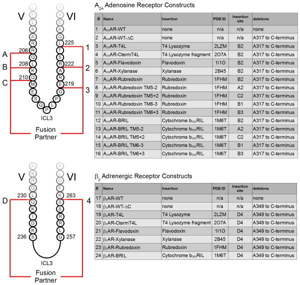
The construct number in the first column as well as the alphanumeric insertion site are used as a reference for all the constructs referred to in the main text. For example, insertion site B2 refers to the initial insertion site of T4L into A2AAR-T4L, between residues L208 and R222.
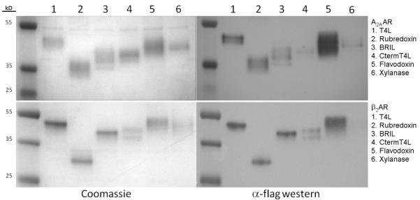
Coomassie stained gels are on the left panels, and the equivalent Western Immunoblots on the right. Multiple receptor bands are in part due to differential glycosylation states of the receptor, and can be consolidated after deglycosylation with PNGaseF and reduction by reducing agent (Figure S1).
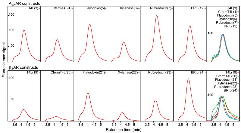
Signals represent fluorescence emission at 350 nm. The rightmost box of each row shows the normalized and overlaid profiles for comparative purposes. Construct numbers are in parenthesis after construct name.
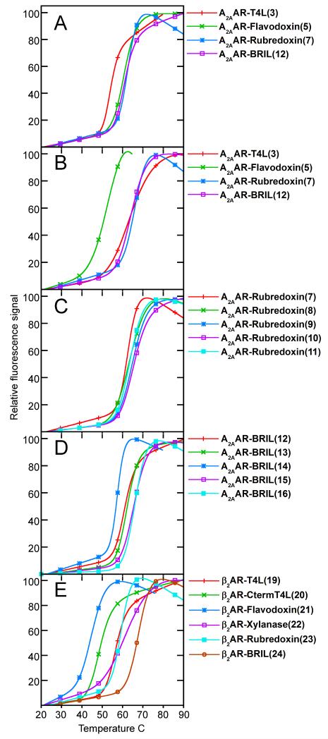
Thermostability profiles for (A) initial A2AAR chimera screen (constructs 3, 5, 7, 12) with ZM241385, (B) initial A2AAR chimera screen (constructs 3, 5, 7, 12) with UK432,097, (C) junction optimization for A2AAR-Rubredoxin with ZM241385 (constructs 7-11), (D) junction optimization for A2AAR-BRIL with ZM241285 (constructs 12-16), and (E) initial β2AR chimera (constructs 19-24) screen with timolol. Construct numbers in parenthesis after name.
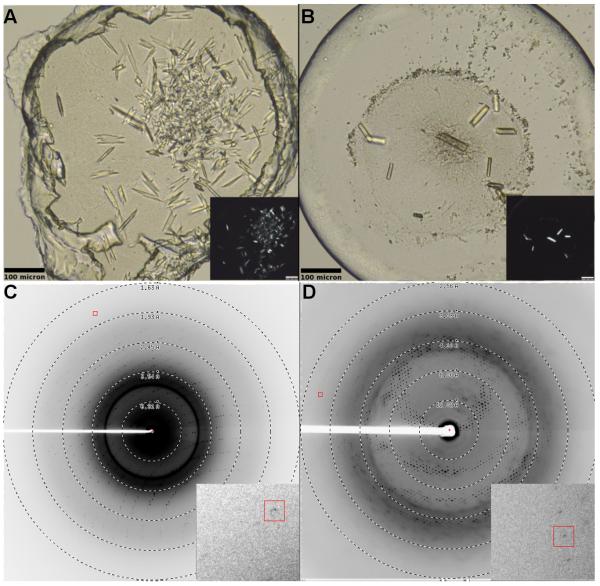
(A) A2AAR-BRIL/ZM241385 (construct 16) and (B) β2AR-BRIL/timolol (construct 24). Crystals grew to approximately 60 × 10 × 3 μm for A2AAR-BRIL and 80 × 15 × 5 μm for β2AR-BRIL. Inset shows the same image under cross polarized light. Diffraction patterns for (C) A2AAR-BRIL and (D) β2AR-BRIL. Inset shows magnified view around diffraction spot enclosed by the red box.
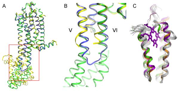
(A) High-resolution crystal structure of A2AAR-BRIL (green) superimposed with A2AAR-T4L (PDB ID 3EML, yellow) and thermostabilized A2AAR (PDB ID 3PWH, blue). (B) Close up view of the junction site enclosed by red box in panel A. The cytoplasmic ends of helix V and helix VI of A2AAR-BRIL and thermostabilized A2AAR superimpose very well, while the helices of A2AAR-T4L must diverge to accommodate the insertion of T4L (T4L domain not shown). (C) Superimposition of the fused BRIL (green) with standalone BRIL (PDB ID 1M6T, orange), cytochrome b562 with heme (PDB ID 256B, magenta) and NMR models of apocytochrome b562 (PDB ID 1YYX, grey) suggest high rigidity in the protein core (bottom half) and high flexibility in the termini and loop2 (top half), especially in the apo structures.
References
-
- Alexandrov AI, Mileni M, Chien EY, Hanson MA, Stevens RC. Microscale fluorescent thermal stability assay for membrane proteins. Structure. 2008;16:351–359. - PubMed
-
- Bjarnadottir TK, Gloriam DE, Hellstrand SH, Kristiansson H, Fredriksson R, Schioth HB. Comprehensive repertoire and phylogenetic analysis of the G protein-coupled receptors in human and mouse. Genomics. 2006;88:263–273. - PubMed
-
- Cherezov V, Peddi A, Muthusubramaniam L, Zheng YF, Caffrey M. A robotic system for crystallizing membrane and soluble proteins in lipidic mesophases. Acta Crystallogr D Biol Crystallogr. 2004;60:1795–1807. - PubMed
Publication types
MeSH terms
Substances
Grants and funding
LinkOut - more resources
Full Text Sources
Other Literature Sources
