

Fluorescein angiography to estimate normal peripheral retinal nonperfusion in children
- PMID: 22681939
- PMCID: PMC3756139
- DOI: 10.1016/j.jaapos.2011.12.157
Fluorescein angiography to estimate normal peripheral retinal nonperfusion in children
Abstract
Purpose: To estimate the normal distance from vascular termini to ora serrata in children's eyes.
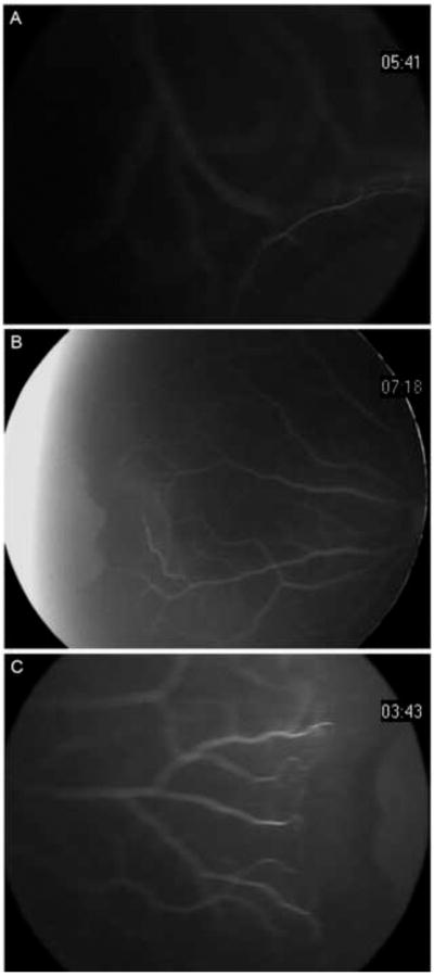
Methods: Clinical records and peripheral fluorescein angiography images of the ora serrata region, taken using scleral indentation and the RetCam system during examination under anesthesia, were retrospectively reviewed from consecutive patients with presumed normal peripheral retinal vasculature. All patients had ocular disease either only in the fellow eye or if in the study eye, to a degree judged not likely to affect peripheral retinal vascular development.
Results: The record review identified 33 eyes of 31 patients with presumed normal peripheral vasculature. Mean age at angiography was 3.8 years (range, 2 months to 13 years). Mean area of nonperfusion was 0.9 disk diameters (DD) temporally (range, 1.5-0.5 DD; SD 0.3) and 0.6 DD nasally (range, 1-0.25 DD; SD 0.2).
Conclusions: In children up to 13 years of age, the avascular retina normally extends 1.5 DD or less temporally and 1.0 DD or less nasally from the ora serrata. Conservatively, ≥ 2 DD of nonperfusion, 3 standard deviations more than normal, should be considered abnormal and a sign of peripheral nonperfusion. These data may serve as preliminary indicators of the range of normal when evaluating diseases with retinal vascular abnormalities in children.
Copyright © 2012 American Association for Pediatric Ophthalmology and Strabismus. Published by Mosby, Inc. All rights reserved.
Figures
References
-
- Rutnin U, Schepens CL. Fundus appearance in normal eyes. II. The standard peripheral fundus and developmental variations. Am J Ophthalmol. 1967;64:840–52. - PubMed
-
- Rutnin U, Schepens CL. Fundus appearance in normal eyes. IV. Retinal breaks and other findings. Am J Ophthalmol. 1967;64:1063–78. - PubMed
-
- Rutnin U, Schepens CL. Fundus appearance in normal eyes. 3. Peripheral degenerations. Am J Ophthalmol. 1967;64:1040–62. - PubMed
-
- Asdourian GK, Goldberg MF. The angiographic pattern of the peripheral retinal vasculature. Arch Ophthalmol. 1979;97:2316–8. - PubMed
-
- Zenker HJ. Fluorescein angiography in inflammation of the peripheral fundus: The normal fluorescein angiographic pattern. I. Ophthalmologica. 1985;190:77–82. - PubMed
MeSH terms
Grants and funding
LinkOut - more resources
Full Text Sources
