. 2012 Jun 9;7(1):13.
doi: 10.1186/1749-8546-7-13.

A combination extract of ginseng, epimedium, polygala, and tuber curcumae increases synaptophysin expression in APPV717I transgenic mice

Affiliations

A combination extract of ginseng, epimedium, polygala, and tuber curcumae increases synaptophysin expression in APPV717I transgenic mice

Jing Shi et al. Chin Med. .

Abstract

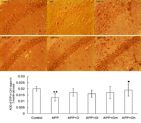
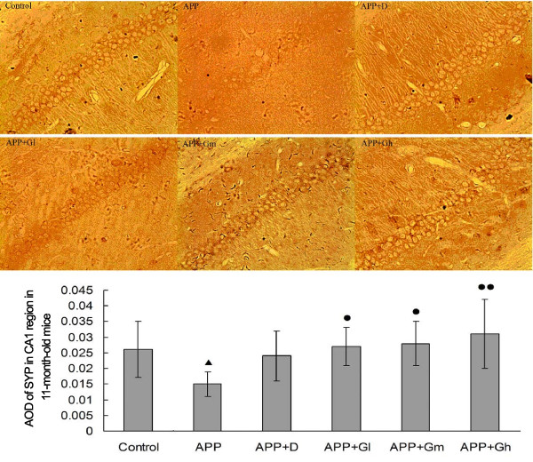
Background: The density of presynaptic markers of synaptic communication and plasticity, especially synaptophysin (SYP), is significantly correlated with cognitive decline and the progression of Alzheimer's disease (AD), indicating that synaptic protection is an important therapeutic strategy for AD. This study aims to investigate the synaptic protective effects of a combination of several active components extracted from the Chinese herbs ginseng, epimedium, polygala and tuber curcumae (GEPT), in the brains of APPV717I transgenic mice.

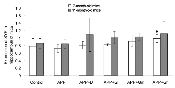
Methods: Three-month-old APPV717I mice were arbitrarily divided into 10 groups (n = 12 per group): APP groups receiving vehicle treatment for four or eight months (model groups), three dose groups of GEPT-treated mice for each treatment period, and donepezil-treated mice for each treatment period. Three-month-old C57BL/6 J mice (n = 12) were also given vehicle for four or eight months (control groups). Vehicle, donepezil or GEPT were intragastrically administered. Immunohistochemistry (IHC) and Western blot analysis were used to assess protein expression in the hippocampal CA1 region and ratios of SYP to β-actin levels in hippocampal tissue homogenate, respectively.

Results: Both IHC and Western blot revealed a decrease in SYP levels in the CA1 region of 7- and 11-month-old APPV717I transgenic mice compared with the control groups, whereas SYP levels were increased in donepezil- and GEPT-treated transgenic mice compared with the APP group. There was a significant difference in the levels of SYP detected by IHC between the GEPT high-dose group and the APP group after 4 months of treatment, and there were significant differences between all three GEPT groups and the APP group after 8 months of treatment. Western blotting showed that the SYP protein-β-actin ratio was decreased in APP mice, while donepezil- and GEPT-treated transgenic mice showed increased trends in the SYP protein-β-actin ratios.

Conclusion: GEPT increases SYP expression and protects synapses before and after the formation of amyloid plaques in the brains of APPV717I transgenic mice.


Figures

Figure 1
Figure 1
Expression of SYP in the hippocampal CA1 region in 7-month-old mice. The effects of GEPT on the expression of SYP in the hippocampal CA1 region in 7-month-old experimental mice were assessed by IHC staining. Average optical densities of the SYP positive neuronal area (anti-body for SYP, 1:400) are expressed as mean ± SD (n = 5). Control: C57BL/6 J mice; APP: APPV717I mice; APP + D: APP mice treated with donepezil; APP + Gl: APP mice treated with GEPT (low dose); APP + Gm: APP mice treated with GEPT (medium dose); APP + Gh: APP mice treated with GEPT (high dose). P < 0.05 vs. APP mice alone, P < 0.01 vs. APP mice; ▴▴P < 0.01 vs. control mice, ANOVA.
Figure 2
Figure 2
Expression of SYP in the hippocampal CA1 region in 11-month-old mice. The effects of GEPT on the expression of SYP in the hippocampal CA1 region in 11-month-old experimental mice were assessed by IHC staining. Average optical densities of the SYP positive neuronal area (anti-body for SYP, 1:400) are expressed as mean ± SD (n = 5). Control: C57BL/6 J mice; APP: APPV717I mice; APP + D: APP mice treated with donepezil; APP + Gl: APP mice treated with GEPT (low dose); APP + Gm: APP mice treated with GEPT (medium dose); APP + Gh: APP mice treated with GEPT (high dose). P <0.05 vs. control group, P < 0.05 vs. APP group, P < 0.01 vs. APP group, one-way ANOVA.
Figure 3
Figure 3
Expression of SYP proteins (SYP–β-actin ratio) in hippocampal tissue homogenates of 7- and 11-month old experimental mice. The effects of GEPT on the expression of SYP in hippocampal tissue homogenates of 7- and 11-month old experimental mice were determined by Western blot (anti-body for SYP, 1:5000) and subsequently analyzed by densitometry. β-actin served as an internal control. Control: C57BL/6 J mice; APP: APPV717I mice; APP + D: APP mice treated with donepezil; APP + Gl: APP mice treated with GEPT (low dose); APP + Gm: APP mice treated with GEPT (medium dose); APP + Gh: APP mice treated with GEPT (high dose). P < 0.05 vs. APP group, one-way ANOVA.

References

    1. Brookmeyer R, Johnson E, Ziegler-Graham K, Arrighi HM. Forecasting the global burden of Alzheimer’s disease. Alzheimers Dement. 2007;3:186–191. doi: 10.1016/j.jalz.2007.04.381. - DOI - PubMed
    1. Selkoe DJ. Normal and abnormal biology of the beta-amyloid precursor protein. Annu Rev Neurosci. 1994;17:489–517. doi: 10.1146/annurev.ne.17.030194.002421. - DOI - PubMed
    1. Selkoe DJ. The molecular pathology of Alzheimer’s disease. Neuron. 1991;6:487–498. doi: 10.1016/0896-6273(91)90052-2. - DOI - PubMed
    1. Hardy J, Higgins G. Alzheimer’s disease: the amyloid cascade hypothesis. Science. 1992;256:184–185. doi: 10.1126/science.1566067. - DOI - PubMed
    1. Davies CA, Mann DMA, Sumpter PQ, Yates PO. A quantitative morphometric analysis of the neuronal and synaptic content of the frontal and temporal cortex in patients with Alzheimer’s disease. J Neurol Sci. 1987;78:151–164. doi: 10.1016/0022-510X(87)90057-8. - DOI - PubMed

LinkOut - more resources