. 2012 Jun 8:11:38.
doi: 10.1186/1476-4598-11-38.

Triplex DNA-binding proteins are associated with clinical outcomes revealed by proteomic measurements in patients with colorectal cancer

Affiliations

Triplex DNA-binding proteins are associated with clinical outcomes revealed by proteomic measurements in patients with colorectal cancer

Laura D Nelson et al. Mol Cancer. .

Abstract

Background: Tri- and tetra-nucleotide repeats in mammalian genomes can induce formation of alternative non-B DNA structures such as triplexes and guanine (G)-quadruplexes. These structures can induce mutagenesis, chromosomal translocations and genomic instability. We wanted to determine if proteins that bind triplex DNA structures are quantitatively or qualitatively different between colorectal tumor and adjacent normal tissue and if this binding activity correlates with patient clinical characteristics.

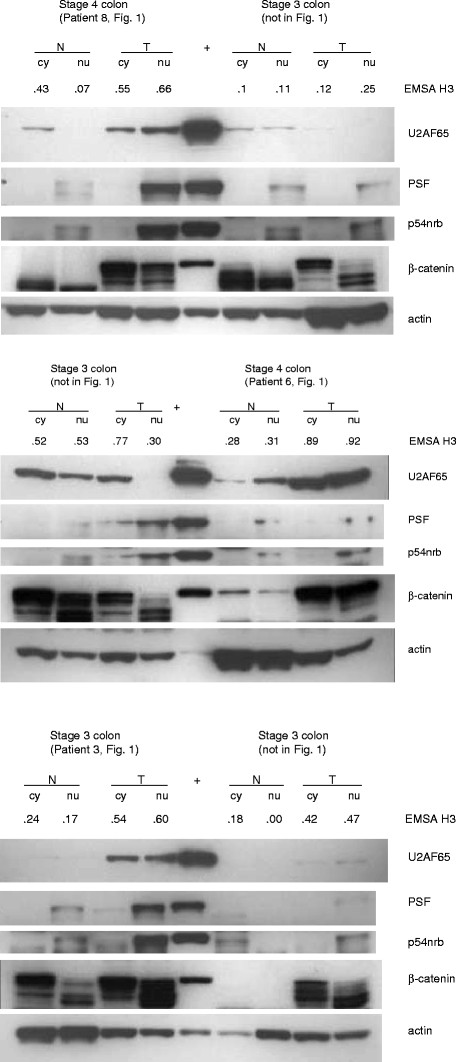
Methods: Extracts from 63 human colorectal tumor and adjacent normal tissues were examined by gel shifts (EMSA) for triplex DNA-binding proteins, which were correlated with clinicopathological tumor characteristics using the Mann-Whitney U, Spearman's rho, Kaplan-Meier and Mantel-Cox log-rank tests. Biotinylated triplex DNA and streptavidin agarose affinity binding were used to purify triplex-binding proteins in RKO cells. Western blotting and reverse-phase protein array were used to measure protein expression in tissue extracts.

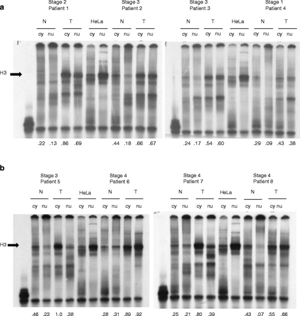
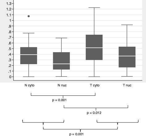
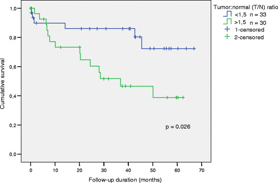
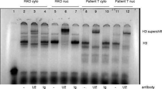
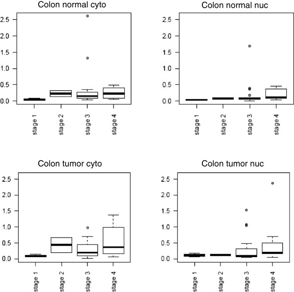
Results: Increased triplex DNA-binding activity in tumor extracts correlated significantly with lymphatic disease, metastasis, and reduced overall survival. We identified three multifunctional splicing factors with biotinylated triplex DNA affinity: U2AF65 in cytoplasmic extracts, and PSF and p54nrb in nuclear extracts. Super-shift EMSA with anti-U2AF65 antibodies produced a shifted band of the major EMSA H3 complex, identifying U2AF65 as the protein present in the major EMSA band. U2AF65 expression correlated significantly with EMSA H3 values in all extracts and was higher in extracts from Stage III/IV vs. Stage I/II colon tumors (p=0.024). EMSA H3 values and U2AF65 expression also correlated significantly with GSK3 beta, beta-catenin, and NF- B p65 expression, whereas p54nrb and PSF expression correlated with c-Myc, cyclin D1, and CDK4. EMSA values and expression of all three splicing factors correlated with ErbB1, mTOR, PTEN, and Stat5. Western blots confirmed that full-length and truncated beta-catenin expression correlated with U2AF65 expression in tumor extracts.

Conclusions: Increased triplex DNA-binding activity in vitro correlates with lymph node disease, metastasis, and reduced overall survival in colorectal cancer, and increased U2AF65 expression is associated with total and truncated beta-catenin expression in high-stage colorectal tumors.


Figures

Figure 1
Figure 1
Electrophoretic Mobility Shift Assay (EMSA) of Resected Tissue Extracts with Purine Triplex DNA. 33P-labeled purine-motif triplex DNA (1 nM) was complexed with 5 μg total protein from normal cytoplasmic (N cy), normal nuclear (N nu), tumor cytoplasmic (T cy) or tumor nuclear (T nu) extracts of tissues obtained from eight selected colorectal cancer patients. 1.25 μg HeLa cytoplasmic and nuclear extracts were used as positive (+) controls. The major EMSA band was H3, indicated with an arrow. Normalized EMSA H3 values are listed below the corresponding samples. The purine-motif triplex probe alone is shown in lane 1.
Figure 2
Figure 2
Normalized EMSA H3 values for all 63 colorectal cancer patient extracts. Box plots indicating the median normalized EMSA H3 values (white lines in the boxes), upper and lower quartiles (25th through 75th percentiles defined by the shaded box), and ranges of data values for each extract type. N cyto, cytoplasmic normal tissue extracts; N nuc, nuclear normal tissue extracts; T cyto, cytoplasmic tumor tissue extracts; Tnuc, nuclear tumor tissue extracts.
Figure 3
Figure 3
Overall Survival according to the tumor:normal (T/N) colorectal tissue nuclear triplex DNA-binding activity ratio. Cut-off = 1.5 (rounded-up median).
Figure 4
Figure 4
Production of a super-shifted H3 band in RKO and patient tissue extracts by super-shift EMSA with a monoclonal antibody against U2AF65. 33P-labeled triplex DNA (1 nM) was complexed with 1.5 μg total protein from RKO cytoplasmic (lanes 2-4), RKO nuclear (lanes 5-7), 5 μg tumor cytoplasm (T cyto lanes 8-10) or tumor nuclear (T nuc lanes 11-12) extracts. Lanes 2, 5, 8, and 11, no antibody; lanes 3, 6, 9, and 12, 400 ng anti-U2AF65 antibody MC3; lanes 4, 7, and 10, mouse IgG antibody (negative control). Each reaction also contained 2 μg poly (dI-dC) carrier DNA. Lane 1, triplex DNA probe alone.
Figure 5
Figure 5
U2AF65 protein expression by colon tumor stage. Total protein (25 μg) from cytoplasmic (cyto) and nuclear (nuc) colon tumor and normal tissue extracts were separated using 10% SDS-PAGE and electro-transferred to nitrocellulose membranes. Blots were incubated with anti-U2AF65 antibody MC-3 and detected using chemiluminescence and autoradiography. Blots were reprobed with an anti-actin antibody, and densitometry was performed using NIH Image J software. U2AF65 expression values were normalized by dividing the actin expression values in each extract, and plotted according to colon tumor stage using the R program (Additional file 6).
Figure 6
Figure 6
Western blots of U2AF65, PSF, p54nrb, and beta-catenin expression in normal and tumor colorectal tissue extracts. Total protein (25 μg) from cytoplasmic (cy) and nuclear (nu) tissue extracts obtained from six selected patients were separated using 10% SDS-PAGE and electro-transferred to nitrocellulose membranes. Blots were incubated with the antibodies against U2AF65, PSF, p54nrb, beta-catenin, and actin, then the appropriate secondary antibody and detected using chemiluminescence and autoradiography. Each patient’s tumor stage and number which are also included in Figure 1, and corresponding EMSA H3 values are shown above the samples.

References

    1. Frank-Kamenetskii MD, Mirkin SM. Triplex DNA structures. Annu Rev Biochem. 1995;64:65–95. doi: 10.1146/annurev.bi.64.070195.000433. - DOI - PubMed
    1. Patel DJ, Phan AT, Kuryavyi V. Human telomere, oncogenic promoter and 5'-UTR G-quadruplexes: diverse higher order DNA and RNA targets for cancer therapeutics. Nucleic Acids Res. 2007;35:7429–7455. doi: 10.1093/nar/gkm711. - DOI - PMC - PubMed
    1. Belotserkovskii BP, De Silva E, Tornaletti S, Wang G, Vasquez KM, Hanawalt PC. A triplex-forming sequence from the human c-MYC promoter interferes with DNA transcription. J Biol Chem. 2007;282:32433–32441. doi: 10.1074/jbc.M704618200. - DOI - PubMed
    1. Nikolova EN, Kim E, Wise AA, O'Brien PJ, Andricioaei I, Al-Hashimi HM. Transient Hoogsteen base pairs in canonical duplex DNA. Nature. 2011;470:498–502. doi: 10.1038/nature09775. - DOI - PMC - PubMed
    1. Hanahan D, Weinberg RA. Hallmarks of cancer: the next generation. Cell. 2011;144:646–674. doi: 10.1016/j.cell.2011.02.013. - DOI - PubMed

Publication types

MeSH terms