Case Reports
. 2012 Jul 13;91(1):15-26.
doi: 10.1016/j.ajhg.2012.05.002. Epub 2012 Jun 7.

TMEM165 deficiency causes a congenital disorder of glycosylation

Affiliations
Case Reports

TMEM165 deficiency causes a congenital disorder of glycosylation

François Foulquier et al. Am J Hum Genet. .

Abstract

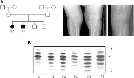
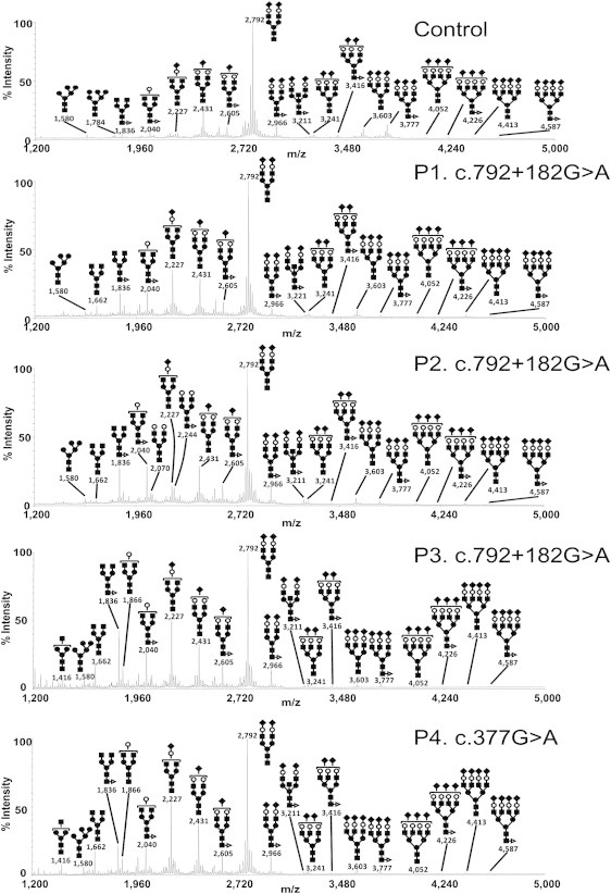
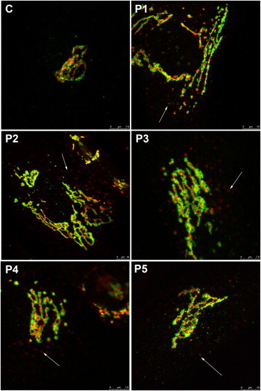
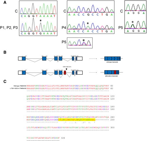
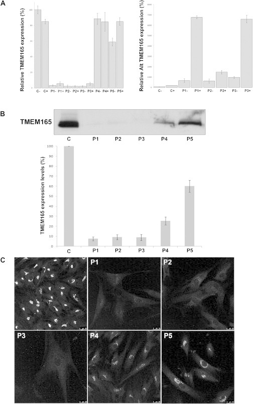
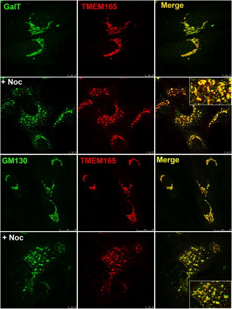
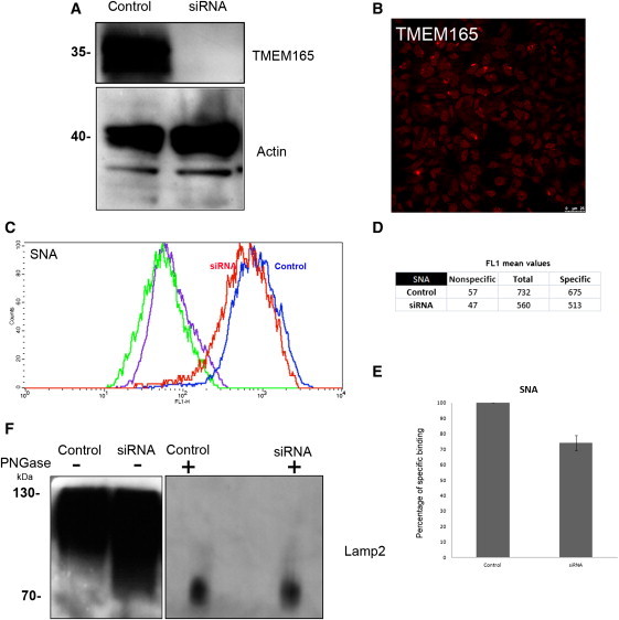
Protein glycosylation is a complex process that depends not only on the activities of several enzymes and transporters but also on a subtle balance between vesicular Golgi trafficking, compartmental pH, and ion homeostasis. Through a combination of autozygosity mapping and expression analysis in two siblings with an abnormal serum-transferrin isoelectric focusing test (type 2) and a peculiar skeletal phenotype with epiphyseal, metaphyseal, and diaphyseal dysplasia, we identified TMEM165 (also named TPARL) as a gene involved in congenital disorders of glycosylation (CDG). The affected individuals are homozygous for a deep intronic splice mutation in TMEM165. In our cohort of unsolved CDG-II cases, we found another individual with the same mutation and two unrelated individuals with missense mutations in TMEM165. TMEM165 encodes a putative transmembrane 324 amino acid protein whose cellular functions are unknown. Using a siRNA strategy, we showed that TMEM165 deficiency causes Golgi glycosylation defects in HEK cells.


Figures

Figure 1
Figure 1
Family tree, Presentation of Radiological Findings, and sTf-IEF Pattern (A) The family of cases 1 and 2 is the index family in this study. Radiological image of a knee in case 1 at the age of 17 years (left panel) and of case 3 at the age of 3.5 years (right panel). Note severe osteopenia, a very thin bone cortex, broad metaphyses, and significant dysplasia of the epiphyses. (B) sTf-IEF pattern from a control and from the individuals under investigation. The number of negative charges is indicated on the right.
Figure 2
Figure 2
Protein N-Glycosylation Deficiencies MALDI-TOF-MS spectra of the permethylated N-glycans from sera of control and TMEM165-deficient individuals. The symbols representing sugar residues are as follows: closed square, N-acetylglucosamine; open circle, mannose; closed circle, galactose; open diamond, sialic acid; and closed triangle, fucose. Linkages between sugar residues have been removed for simplicity.
Figure 3
Figure 3
Alterations in Golgi Structure Golgi localization of GM130 (green) and TGN46 (red) in control and TMEM165-deficient fibroblasts. The cells were double labeled with antibodies against GM130 and TGN46 and visualized by confocal microscopy. The arrows point to fragmented Golgi.
Figure 4
Figure 4
TMEM165 Mutations (A) In the left panel is the sequence alignment of the genomic DNA fragment from a control C and cases P1, P2, and P3 homozygous for the G>A transition at position c.792+182. In the middle and right panels is the sequence alignment of the cDNA fragment from a control C and cases P4 and P5. P4 is homozygous for the G>A transition at position c.377, and P5 is compound heterozygous for the C>T transition at position 376 and the G>A transition at position 910. Nucleotides affected by the mutations and the predicted effects on protein length are indicated above each sequence. (B) Schematic representation of the predicted genomic (top) and cDNA (bottom) structure of TMEM165 in the control and cases P1, P2, and P3. Coding exons are shown in blue boxes, and introns (not drawn to scale) are represented by lines. The beginning and end of the open reading frame are labeled; the numbering above each exon is based on coding nucleotides. The red box represents the alternative exon 4 (4′) used in cases P1, P2, and P3. (C) Predicted effect of the c.792+182G>A mutation on TMEM165. Sequence alignment was compared between wild-type TMEM165 and mutant TMEM165. The amino acids that changed as a result of the mutation are highlighted in yellow. The mutated TMEM165 is truncated and shortened by 94 amino acids.
Figure 5
Figure 5
Functional Analyses of TMEM165 Mutations (A) Quantification of the wild-type (left panel) and alternative (alt) TMEM165 transcripts (right panel) in controls and affected individuals by qPCR with (+) and without (−) puromycin. Expression is measured relative to a housekeeping gene. Values plotted with a wild-type control group untreated with puromycin are set to 1. Error bars represent ± SEM. Expression of TMEM165 in control and TMEM165-deficient fibroblasts: steady-state levels of expression (B) and intracellular distribution (C). (B) Immunoblots of whole-cell lysates from control (C) and TMEM165-deficient (P) fibroblasts. Actin levels were used as a loading control. (C) Control and TMEM165-deficient fibroblasts were processed for immunofluorescence microscopy with TMEM165 antibodies. For comparison, images were collected under identical settings.
Figure 6
Figure 6
Subcellular Localization of Wild-Type TMEM165 Indirect double immunofluorescence staining of control fibroblasts treated in the absence and presence of nocodazole (+ Noc). The cells were then double labeled with antibodies against TMEM165 and with antibodies against either GalT or GM130. The cells were then examined by confocal microscopy. In the presence of nocodazole, close examination of the overlays shows that the colocalization of TMEM165 with GalT is more complete than it is with GM130.
Figure 7
Figure 7
Depletion of TMEM165 Slightly Affects Golgi Glycosylation (A) HEK cells were transfected with nontarget siRNA (“Control”) or siRNA targeted to TMEM165 (“siRNA”). Seven days after transfection, cells were lysed and equivalent amounts of homogenate were subjected to SDS PAGE and immunoblotted with antibodies against TMEM165 and actin. (B) Indirect immunofluorescence staining of the siRNA TMEM165 HEK cells with the use of anti-TMEM165. (C) HEK cells were transfected with nontarget siRNA (“Control”) or siRNA targeted to TMEM165 (“siRNA”). Seven days after transfection, cells were harvested, permeabilized, and incubated with FITC-labeled SNA for flow-cytometry analysis. Histogram plots of control (blue line) and siRNA-TMEM165-transfected (red line) HEK cells analyzed for SNA binding and incubated with FITC-labeled SNA are shown. For each condition, the fluorescence peak corresponding to SNA binding in the presence of 200 mM lactose is represented (nonspecific binding: green line for control and violet line for siRNA). Specific binding was calculated as the difference between the mean fluorescence intensities of total and nonspecific binding peaks (see D). (E) The results were expressed as the percentages of specific lectin binding to cells. In the calculation, specific lectin binding to control cells, which corresponds to the difference between the total and the nonspecific-binding peaks, was considered as 100%. (F) HEK cells were transfected with nontarget siRNA (“Control”) or siRNA targeted to TMEM165 (“siRNA”). Seven days after transfection, cells were lysed (incubated or not with PNGase [1,000 U per sample]), and equivalent amounts of homogenate were subjected to SDS and immunoblotted with antibodies against Lamp2.

References

    1. Jaeken J., Matthijs G. Congenital disorders of glycosylation: A rapidly expanding disease family. Annu. Rev. Genomics Hum. Genet. 2007;8:261–278. - PubMed
    1. Jaeken J., Hennet T., Matthijs G., Freeze H.H. CDG nomenclature: Time for a change! Biochim. Biophys. Acta. 2009;1792:825–826. - PMC - PubMed
    1. Schachter H., Freeze H.H. Glycosylation diseases: Quo vadis? Biochim. Biophys. Acta. 2009;1792:925–930. - PMC - PubMed
    1. Foulquier F. COG defects, birth and rise! Biochim. Biophys. Acta. 2009;1792:896–902. - PubMed
    1. Wu X., Steet R.A., Bohorov O., Bakker J., Newell J., Krieger M., Spaapen L., Kornfeld S., Freeze H.H. Mutation of the COG complex subunit gene COG7 causes a lethal congenital disorder. Nat. Med. 2004;10:518–523. - PubMed

Publication types