Comparative Study
. 2012 Jun 29;36(6):1003-16.
doi: 10.1016/j.immuni.2012.05.017. Epub 2012 Jun 7.

Positive and negative signaling through SLAM receptors regulate synapse organization and thresholds of cytolysis

Affiliations
Comparative Study

Positive and negative signaling through SLAM receptors regulate synapse organization and thresholds of cytolysis

Fang Zhao et al. Immunity. .

Abstract

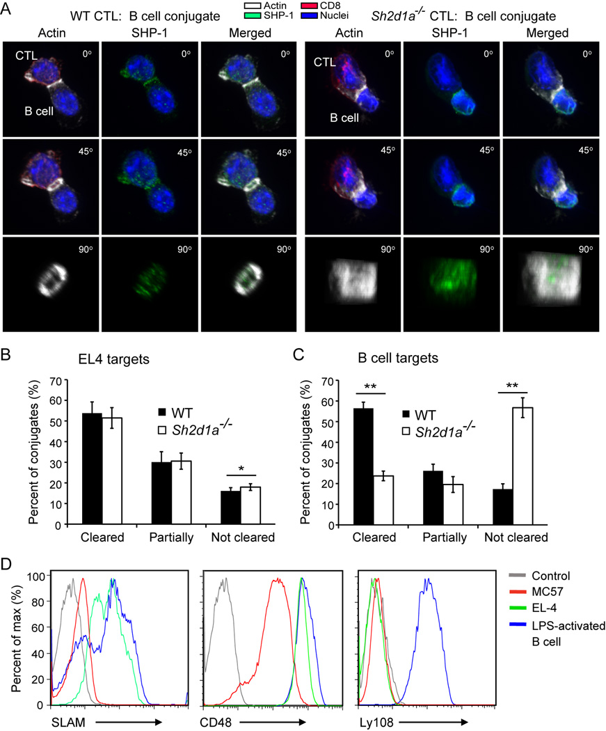
X-linked lymphoproliferative syndrome, characterized by fatal responses to Epstein-Barr virus infection, is caused by mutations affecting the adaptor SAP, which links SLAM family receptors to downstream signaling. Although cytotoxic defects in SAP-deficient T cells are documented, the mechanism remains unclear. We show that SAP-deficient murine CD8(+) T cells exhibited normal cytotoxicity against fibrosarcoma targets, yet had impaired adhesion to and killing of B cell and low-avidity T cell targets. SAP-deficient cytotoxic lymphocytes showed specific defects in immunological synapse organization with these targets, resulting in inefficient actin clearance. In the absence of SAP, signaling through the SLAM family members Ly108 and 2B4 resulted in increased recruitment of the SHP-1 phosphatase, associated with altered SHP-1 localization and decreased activation of Src kinases at the synapse. Hence, SAP and SLAM receptors regulate positive and negative signals required for organizing the T cell:B cell synapse and setting thresholds for cytotoxicity against distinct cellular targets.


Figures

Figure 1
Figure 1. SAP-deficient CTLs have a selective defect in cytolysis of and adhesion to B cell and low avidity T cell targets
(A) Cytolysis of MC57, EL4 and LPS-activated WT B cell targets, pulsed with 1 µM OVA257–264, by WT and Sh2d1a−/− CTLs. (B) Intracellular IFN-γ and IL-2 production by WT and Sh2d1a−/− CTLs after stimulation with OVA257–264 pulsed B cells. Representative of 3 independent experiments. (C–D) Cytolysis of MC57 and EL4 targets pulsed with 30 nM OVA257–264 (C) or 1 µM G4 APL (D) by WT and Sh2d1a−/− CTLs. Graphs show the average cytotoxicity from triplicate wells ±SD for varying effector to target (E:T) ratios, representative of 4 or more experiments. (E–F) Adhesion of WT and Sh2d1a−/− CTLs to WT B cells (E) or EL4 cells (F), pulsed with the indicated concentrations of OVA257–264 or the G4 APL. Graphs show the average of triplicates ±SD, representative of 3 independent experiments.
Figure 2
Figure 2. SAP-deficient CTLs exhibit abnormal actin organization and reduced centrosome docking at the T:B cell IS
(A) LPS-activated WT B cell targets, pulsed with 1 µM OVA257–264, conjugated with WT or Sh2d1a−/− CTLs, examined from the side, as confocal projections in the x–y plane (top row), or at 45° (middle row) and 90° (en face, bottom row) rotations in the y–z plane. Scale bars represent either 10 µm (top and middle row) or 5 µm (bottom row). Nuclei (blue), CD8 (red), talin (green), and actin (white). (B–D) Quantification of actin localization at the IS with WT B cell targets pulsed with 1 µM OVA257–264 peptide (B), EL4 targets pulsed with 30 nM OVA257–264 peptide (C), or EL4 targets pulsed with 1 µM OVA257–264 peptide (D). (E) Centrosome localization, shown as confocal projections in the x–y plane. Nuclei (blue), CD8 (white), Lck (green), and γ-tubulin (red). (F–G) Quantification of centrosome positioning in WT and Sh2d1a−/− CTL conjugates with EL4 (F) or WT B cell (G) targets pulsed with 1 µM OVA257–264 peptide. (H–J) Quantification of centrosome localization in WT and Sh2d1a−/− CTLs exhibiting the phenotypes of actin cleared (H), partially cleared (I), or not cleared (J) at the IS with WT B cell targets pulsed with 1 µM OVA257–264 peptide. Data shown are averages of 3 or more experiments ±SEM, with >45 conjugates scored per genotype for each experiment. *p < 0.05, **p < 0.005, as determined by Paired Student’s T-tests.
Figure 3
Figure 3. SAP-deficient CTLs exhibit abnormal SHP-1 accumulation at the T:B cell IS
(A) WT and Sh2d1a−/− CTL conjugates with LPS-activated WT B cells, examined as in Figure 2A. Nuclei (blue), CD8 (red), SHP-1 (green), and actin (white). (B–C) Quantification of SHP-1 localization at the IS of WT and Sh2d1a−/− CTL conjugates with EL4 (B) or LPS-activated WT B cell (C) targets pulsed with 1 µM OVA257–264 peptide. Data depicted are the average of 3 or more experiments ±SEM, with 35–60 conjugates scored per genotype for each experiment. **p < 0.005, as determined by Paired Student’s T-tests. (D) Expression of SLAM, CD48 and Ly108 on MC57, EL4 and LPS-activated WT B cell targets.
Figure 4
Figure 4. Ly108 and 2B4 show increased association with SHP-1 in the absence of SAP: requirement for Ly108 intracellular tyrosines
(A–B) Lysates from WT and Sh2d1a−/− CTLs, stimulated with WT B cells (NP, no peptide, or 1 µg/mL OVA257–264 pulsed), were immunoprecipitated for Ly108 (A) or 2B4 (B) and immunoblotted for SHP-1 and Ly108 or 2B4, respectively. Representative immunoblots shown, from 3 or more independent experiments. TCL, total cell lysate. (C–E) Slamf6−/− and Slamf6−/−Sh2d1a−/− CTLs, transfected with either WTLy108-GFP (C) or Ly108-AllF-GFP (D), were conjugated with WT B cell targets and shown as confocal projections in the x-y plane (top row) or en face (lower row). Nuclei (blue) and SHP-1 (red). (E) Quantification of SHP-1 localization at the T:B cell IS of Slamf6−/− and Slamf6−/−Sh2d1a−/− CTLs transfected with WTLy108-GFP or Ly108-AllF-GFP. Data shown are representative of two experiments. (F) Slamf6−/− and Slamf6−/−Sh2d1a−/− CTLs, transduced with retroviral vectors expressing either Ly108 or Ly108-AllF, were stimulated with 1 µg/mL OVA257–264 pulsed WT B cells. Lysates were immunoprecipitated for Ly108 and immunoblotted for SHP-1 and Ly108. Representative immunoblot shown from 2 independent experiments.
Figure 5
Figure 5. SAP-deficient CTLs exhibit abnormal p-Src localization and decreased tyrosine phosphorylation at the T:B cell IS and reduced Vav1 phosphorylation
(A) Representative immunofluorescence images of p-Src (pY416) localization, shown as en face reconstructions: p-Src (green) and actin (white). Scale bars represent 5 µm. (B–C) Quantification of p-Src (pY416) localization at the IS of WT and Sh2d1a−/− CTL conjugates with EL4 (B) or WT B cell (C) targets pulsed with 1 µM OVA257–264 peptide. Data are the average of 3 experiments ±SEM, with over 40 conjugates scored per genotype for each experiment. *p < 0.05, **p < 0.005, as determined by Paired Student’s T-tests. (D) Mean intensity of pTyr staining at the IS of WT and Sh2d1a−/− CTL conjugates with EL4 or WT B cell targets pulsed with 1 µM OVA257–264 peptide. Data shown is representative of 3 independent experiments. *p < 0.0001, as determined by Student’s T-tests. (E) Lysates from WT and Sh2d1a−/− CTLs, stimulated with 1 µg/ml OVA257–264 pulsed WT B cell targets, were immunoprecipitated for Vav1 and blotted for phosphotyrosine (4G10) and Vav1. Representative immunoblot shown from 3 independent experiments.
Figure 6
Figure 6. Negative Signaling through the SLAM family receptors is mediated through SHP-1
(A–B) WT and Sh2d1a−/− CTLs were serum-starved and pre-incubated with the SHP-1/2 inhibitor NSC-87877 for 2 hr at 37°C, before examination of cytotoxicity against WT B cell targets pulsed with 1 µM OVA257–264 peptide (A) or EL4 targets pulsed with 1 µM G4 APL (B). Data shown are the average of triplicate wells ±SD for a range of effector to target (E:T) ratios. Representative of 3 independent experiments.
Figure 7
Figure 7. SLAM family receptor engagement is required for the phenotype of SAP-deficient CTLs
(A–B) Cytotoxicity (left) and actin organization at the IS (right) of WT and Sh2d1a−/− CTLs against Slamf6−/− B cells (A) or Ly108-expressing EL4 targets (B) pulsed with 1 µM OVA257–264 peptide. Quantifications of actin organization depict the average of 3 experiments ±SEM and represent more than 40 conjugates per genotype for each experiment. *p < 0.05 as determined by Paired Student’s T-tests. (C) Adhesion of WT, Sh2d1a−/−, Slamf6−/−, and Slamf6−/−Sh2d1a−/− CTLs to WT or Slamf6−/− B cells, pulsed with the indicated concentrations of OVA257–264 or the G4 APL. Graphs show the average of 3 independent experiments. *p < 0.05 as determined by Paired Student’s T-tests. (D–E) Cytotoxicity of WT CTLs against WT or Slamf6−/− B cells (D), or of WT and Slamf6−/− CTLs against control or Ly108-expressing EL4 targets (E). Cytolysis shown is an average of triplicate wells ±SD for a range of effector to target (E:T) ratios, representative of 3 or more experiments. (F) Lysates from WT and Slamf6−/− T cells, stimulated by cross-linking antibodies against anti-CD3 alone or anti-CD3 and anti-Ly108, were immunoblotted for ERK phosphorylation. Representative immunoblot shown from 3 independent experiments.

Similar articles

Cited by

References

    1. Beal AM, Anikeeva N, Varma R, Cameron TO, Vasiliver-Shamis G, Norris PJ, Dustin ML, Sykulev Y. Kinetics of early T cell receptor signaling regulate the pathway of lytic granule delivery to the secretory domain. Immunity. 2009;31:632–642. - PMC - PubMed
    1. Booth C, Gilmour KC, Veys P, Gennery AR, Slatter MA, Chapel H, Heath PT, Steward CG, Smith O, O'Meara A, et al. X-linked lymphoproliferative disease due to SAP/SH2D1A deficiency: a multicenter study on the manifestations, management and outcome of the disease. Blood. 2011;117:53–62. - PMC - PubMed
    1. Bottino C, Falco M, Parolini S, Marcenaro E, Augugliaro R, Sivori S, Landi E, Biassoni R, Notarangelo LD, Moretta L, Moretta A. NTB-A, a novel SH2D1A-associated surface molecule contributing to the inability of natural killer cells to kill Epstein-Barr virus-infected B cells in X-linked lymphoproliferative disease. J Exp Med. 2001;194:235–246. - PMC - PubMed
    1. Brossard C, Feuillet V, Schmitt A, Randriamampita C, Romao M, Raposo G, Trautmann A. Multifocal structure of the T cell - dendritic cell synapse. Europ J Immunol. 2005;35:1741–1753. - PubMed
    1. Campi G, Varma R, Dustin ML. Actin and agonist MHC-peptide complex-dependent T cell receptor microclusters as scaffolds for signaling. J Exp Med. 2005;202:1031–1036. - PMC - PubMed

Publication types

MeSH terms

Substances